Vascular diseases. Vascular diseases, pathology with vascular factor Damage to the blood vessels of the brain is called
The word "aneurysm" comes from the Latin "aneurysma", which means dilation. An aneurysm is an abnormal local enlargement of the wall of a blood vessel, usually an artery, due to a defect, disease or injury.
Aneurysms can be true or false. A false aneurysm is a cavity filled with a blood clot. There are two types of true intracranial aneurysms: saccular and fusiform (fusiform).
Types of cerebral aneurysms
Common causes of intracranial aneurysms are hemodynamically induced or degenerative vascular damage, atherosclerosis (usually leading to the formation of fusiform aneurysms), and concomitant vasculopathy (for example, fibromuscular dysplasia). Rare causes include trauma, infection, certain substances, and tumors (primary or metastatic).
Saccular aneurysms are round, berry-like protrusions that usually develop at the bifurcation of an artery, most often in the area of the arterial circle of the brain (Circle of Willis). These are true aneurysms, that is, dilatation of the vascular wall occurs due to the weakness of all its layers.
The normal arterial wall consists of three layers: intima (represented by the endothelium - the innermost layer); The media, which consists of smooth muscle and adventitia, is the outermost layer consisting of connective tissue. The aneurysmal sac itself usually consists of only two layers - intima and adventitia. The intima is usually normal; however, subintimal cell proliferation may be detected. The internal elastic membrane is usually thinned or completely absent, and the media ends at the point where the neck of the aneurysmal sac forms in close proximity to the supporting vessel.
Etiology
Most saccular intracranial aneurysms were previously thought to be the result of a gradual protrusion in the area of a congenital defect of the vascular wall that develops throughout life.
Current research has found evidence of congenital, acquired, and hereditary arterial wall defects. Despite the existence of genetic syndromes accompanied by the formation of aneurysms, most of them most likely develop due to hemodynamic and degenerative damage to the vascular wall. The prevalence, growth, presence of blood clots in the cavities and even rupture of aneurysms can be explained using hemodynamic theory.
Less common causes of saccular aneurysms are trauma, tumors, and drugs (cocaine).
Prevalence
The true prevalence of intracranial aneurysms is unknown, but is thought to range from 1 to 6% of the entire population. Published data vary depending on what is considered an aneurysm and depending on what material was studied (autopsy data or angiographic data). In one study of patients undergoing angiography, incidental aneurysms were found in 5.6% of cases, and in another in 1% of patients undergoing panangiography for subarachnoid hemorrhage. A family history of intracranial aneurysms has also been described.
Accompanying illnesses
Congenital anomalies of intracranial vessels, such as fenestration of the vertebrobasilar joint or the presence of a persistent trigeminal artery, are associated with an increased incidence of saccular aneurysms. Aneurysms in combination with fenestration were found both on the side of the fenestration and on the opposite side.
Vasculopathies such as fibromuscular dysplasia, connective tissue diseases, and spontaneous arterial dissection are also associated with an increased risk of aneurysm formation.
Diseases in which there is an increased risk of cerebral aneurysms:
- Polycystic kidney disease
- Coarctation of the aorta
- Abnormal vessels
- Fibromuscular dysplasia
- Connective tissue diseases (Marfan syndrome, Ehlers-Danlos syndrome)
- Presence of vascular malformations and fistulas in other organs
Plurality
Multiple intracranial aneurysms occur in 10 - 30%. About 75% of patients with multiple aneurysms have two aneurysms, 15% have three, and 10% have more than three. Multiple aneurysms are more common in women. The male to female ratio for multiple aneurysms is 5:1, and for three or more aneurysms it is 11:1.
Multiple aneurysms also occur in vasculopathies.
Multiple aneurysms can be bilaterally symmetrical (mirror aneurysms) or localized symmetrically on different vessels. There may be several aneurysms on one artery.

Age of onset
Aneurysms typically become symptomatic in people aged 40–60 years, with a peak incidence of SAH (subarachnoid hemorrhage) at ages 55–60 years. In children, intracranial aneurysms are rare and account for no more than 2% of all cases. Aneurysms in pediatrics most often occur after injuries or mycoses, and somewhat more often in boys. Aneurysms found in children are also slightly larger than aneurysms found in adults, with an average diameter of 17 mm.
Localization and clinical signs of aneurysms
Aneurysms often develop in the area of bifurcations of the main arteries of the brain. Most saccular aneurysms occur in the area of the circle of Willis or the bifurcation of the middle cerebral artery (MCA).
Aneurysms of the anterior sections of the circle of Willis: Approximately 86.5% of all intracranial aneurysms occur in this section of the arterial circle. Common sites of aneurysms include the anterior communicating artery (ACA) -30%, the internal carotid artery (ICA) at the origin of the posterior communicating artery (25%), and the MCA bifurcation (20%). Aneurysm of the bifurcation of the ICA is observed in 7.5%, and of the pericallosal/callosomarginal artery in 4%.
Posterior circle of Willis aneurysms: account for about 10% of all cerebral aneurysms. 7% are aneurysms of the bifurcation of the basilar artery, and the remaining 3% are aneurysms of the posterior inferior cerebellar artery (PICA) at its origin from the vertebral artery (VA).
More rare localizations of aneurysms: account for 3.5% of all aneurysms. This includes aneurysms of the superior cerebellar artery and anterior inferior cerebellar artery at their origin from the main artery. Saccular aneurysms of these localizations are quite rare.
Clinical signs
Most aneurysms do not cause clinical symptoms until they rupture, which carries a high risk of death.
Subarachnoid hemorrhage
The most common complication of aneurysms is non-traumatic subarachnoid hemorrhage (SAH). In Russia, 80-90% of non-traumatic SAH occur due to rupture of intracranial aneurysms. Another 5% are associated with AVMs (arteriovenous malformations) or tumors and the remaining 5-15% are idiopathic.
When an aneurysm ruptures, the patient usually experiences a sharp, severe headache, which is often compared to a blow. The presence of meningeal syndrome confirms the possible diagnosis of SAH. Subhyaloid hemorrhages, often bilateral, which are localized between the retina and the vitreous, can be observed in 25% of cases.  The most widely used scale for assessing the severity of clinical symptoms of hemorrhage is the Hunt-Hess scale, which also correlates with disease outcome.
Clinical symptoms of aneurysms not associated with hemorrhages are quite rare. Some intracranial aneurysms are accompanied by cranial nerve neuropathy. The most striking example is neuropathy of the third cranial nerve (oculomotor) with aneurysms of the posterior communicating artery. Other, more rare symptoms are visual field disturbances in ophthalmic ICA aneurysms, causing compression of the optic nerves, seizures, headache, transient ischemic attacks due to secondary embolism (usually found in giant, partially thrombosed MCA aneurysms). So-called giant aneurysms (diameter >2.5 cm) more often present with various neurological symptoms due to their mass effect.
Diagnosis of aneurysms
Three main techniques are used to determine the size, location, and morphology of intracranial aneurysms: contrast-enhanced computed tomography (CT angiography), MRI, and cerebral puncture angiography. The preferred methods for screening diagnosis of unruptured aneurysms are MRI and CT agnography, while needle angiography is the method of choice in patients who have undergone SAH.
Needle angiography
 This method continues to be the standard for determining the basic characteristics of intracranial aneurysms. The currently existing methods of selective three-dimensional puncture angiography have significantly expanded the possibilities of studying the anatomy of aneurysms. This technique, introduced in the late 90s, is now widely used in many clinics. Images can be examined from any angle, providing a more detailed view of aneurysm anatomy compared to 2D images.
The role of cerebral angiography in patients with nontraumatic SAH is to identify any aneurysm, determine its relationship with the supporting vessel and adjacent arteries, identify vascular spasm, and most importantly, determine which treatment option is most appropriate for the patient.
Technically correct cerebral angiography is considered the most important and most accurate method for diagnosing SAH, but many authors have reported the successful use of CT angiography.
CT scan
Aneurysms that are quite large in size (usually more than 10 mm) or aneurysms containing calcifications in their cavity can be visualized using non-contrast CT. During the examination, erosions of the bones of the base of the skull may be detected at the site of large aneurysms.
When performing non-contrast CT, typical non-thrombosed aneurysms are visible as well-circumscribed isodense or slightly hyperdense formations localized in the suprasellar arachnoid space or in the region of the Sylvian fissure. Aneurysms are well contrasted after administration of a contrast agent. Angiographic-like images of the cerebral vessels can be obtained by rapidly injecting contrast and simultaneously performing a thin-section CT scan (called CT angiography). Various 3D image processing techniques allow you to obtain fairly clear and detailed images. Such studies allow solving many problems, including a detailed assessment of the relationship between the aneurysm and surrounding structures.
The accuracy of high-resolution CT angiography in diagnosing aneurysms with a diameter of 3 mm or more reaches 97%. 
 Detecting aneurysms using MRI can be challenging. The MR signal depends on the presence and direction of blood flow in the aneurysm, on the presence of blood clots, fibrosis and calcifications.
Aneurysms can produce either high or low signal intensity on routine MRI, depending on the characteristics listed and the pulse sequence used. Typical fast-flowing aneurysms appear as well-circumscribed lesions demonstrating high-speed signal loss on T1- and T2-weighted images. Some signal heterogeneity may be due to the presence of turbulent flows in the aneurysm cavity. Intravenous administration of contrast medium does not usually lead to its accumulation in the cavity of aneurysms with high blood flow, but its accumulation in the walls of the aneurysm can be observed.
The macroscopic motion of spins in moving blood, simultaneously with the suppression of the signal from stationary tissues, can be used to obtain images of brain vessels. These images can be assessed individually or as MR angiograms .
Treatment of cerebral aneurysms
1. Surgical clipping
The goal of surgery is usually to apply a special clip to the neck of the aneurysm to exclude the aneurysm from the bloodstream without occluding a normal vessel. When an aneurysm cannot be clipped due to its characteristics or the patient's serious condition, the following alternative interventions can be performed:
Envelopment: Although envelopment should never be the goal of surgery, circumstances may force the surgeon to resort to this method (eg, in fusiform basilar artery aneurysms). Plastic rubber may be better in this situation than muscle or gauze. Envelopment can be done with silk or muslin, with muscle, or with plastic or other polymer. Some studies show better results when using plastic, but others indicate no difference when using natural fabrics.
Tripping: the effectiveness of the procedure requires clipping or ligation (ligation) of the proximal and distal sections of the carrier artery. The operation can be supplemented by the application of intra-extracranial anastomoses to ensure blood flow distal to the ligated segment of the artery.
Proximal ligation: Proximal ligation has been used with some success for giant aneurysms, usually located in the vertebrobasilar territory. Currently, it is preferable to use existing endovascular treatment methods for aneurysms in these locations.
After craniotomy (craniotomy), microsurgical techniques are used under microscope control to free the neck of the aneurysm from the feeding vessels without causing aneurysm rupture. The operation ends with clipping of the neck of the aneurysm to prevent blood flow in it. There are clips of various types, configurations, sizes and lengths that are MRI compatible.
Intraoperative complications and mortality associated with clipping surgery depend on the history of previous rupture. Ruptured aneurysms are more difficult to treat surgically and the incidence of complications is higher. In surgical treatment of unruptured aneurysms, the complication rate is approximately 4-10.9%, and the mortality rate is 1-3%.
2. Endovascular treatment
The technique of endovascular treatment of aneurysms has become widespread over the past 15 years. Initially, the method consisted of embolization of the feeding vessel with a balloon. The procedure was soon replaced by direct obliteration of the aneurysm lumen, first with a detachable balloon and then with microcoils. Separation usually occurs 2-10 minutes after satisfactory placement of the catheter in the neck of the aneurysm.
 A
A  B
B 
Endovascular treatment of aneurysms (A - diagram, B - contrast-enhanced aneurysm of the fork of the main artery, C - after introducing 6 coils into the aneurysm cavity, the aneurysm is turned off from the bloodstream)
Aneurysm obliteration with coils is becoming the preferred treatment for aneurysms in many centers. While embolization was originally used only for aneurysms that were not accessible to direct surgical intervention, this technique is now applicable to most aneurysms.
Arteriovenous malformations of the brain
 AVMs are congenital anomalies consisting of a complex tangle of arteries and veins connected by one or more fistulas. The vascular conglomerate is called an AVM node. The node does not have a capillary bed, and the supplying arteries drain directly into the veins. The arteries have an underdeveloped muscle layer. Draining veins are often dilated due to high blood flow through the fistula. The process of formation of abnormal vessels still remains unknown.
AVMs cause neurological symptoms through three mechanisms. The first of these is hemorrhages, which can be subarachnoid, intraventricular or, most often, parenchymal. Second, even in the absence of hemorrhage, AVMs can manifest as seizures. Approximately 15 - 40% of patients present with seizures. Finally, neurological deficits that progress over several months or years can develop in 6–12% of patients. This slowly progressive neurological deficit develops due to a lack of blood flow to functionally significant areas of the brain surrounding the AVM (the so-called steal phenomenon). In some cases, neurological deficits may be caused by the mass effect of an enlarging AVM or venous hypertension in the draining veins.
Frequency of occurrence
Globally, the incidence of AVM ranges from 0.89 to 1.24 per 100,000 population per year, based on reports from Australia, Sweden and Scotland. In Scotland, the incidence of AVM reaches 18 per 100,000 population per year.
In the United States, the prevalence of AVM, according to a prospective study, was 1.34 per 100,000 population per year.
Morbidity and mortality
Although 300,000 patients in the United States alone have identified AVMs, only 12% of them become symptomatic. Death occurs in 10-15% of patients who develop hemorrhage.
1) Hemorrhage. In a population-based study, 38–70% of all AVMs present with hemorrhage. The overall risk of developing hemorrhage in patients with identified AVMs is approximately 2-4% per year. Patients with previous hemorrhage are at increased risk of developing recurrent hemorrhages, especially during the first year after the first episode. The incidence of hemorrhagic complications increases progressively after the first year from the onset of the disease. Clinical and angiographic features associated with a high risk of recurrent hemorrhage include male sex of the patient, small AVM size, location in the basal ganglia and posterior fossa, drainage into the deep veins of the brain, one or a small number of draining veins, high supply pressure arteries, measured by angiography.
Table 1. Risk of hemorrhage from AVM
2) Seizures and symptomatic epilepsy. Seizures not associated with hemorrhages develop in 15-40% of patients with cerebral AVMs. They can be local or generalized. Satisfactory control of seizures can usually be achieved with conventional anticonvulsants. Convulsive manifestations are usually observed in young patients, with large AVMs, lobar AVM localization (especially in the temporal lobe), and with AVMs feeding from the middle cerebral artery.
3) Headache and migraine. In the general population, headache due to AVM is a very unusual symptom. Headache not associated with hemorrhage occurs in 4-14% of patients and can be persistent. It may be migraine-like or less specific and more generalized.
Diagnosis of AVM
Computed tomography (CT)- During this test, X-rays are used to obtain flat or 3D images of the brain. A CT scan can detect the presence of hemorrhage, which indicates the presence of an AVM.

Magnetic resonance imaging (MRI)- Using a strong magnetic field, MRI allows you to generate flat or 3D images of the brain and diagnose the presence of any vascular pathology. The MR angiography procedure helps to obtain a detailed image of the blood vessels in the brain. The procedure is painless and does not cause damage to human tissue.

Angiography is a special x-ray procedure that shows the exact anatomical structure of the patient's brain vessels and is extremely important in the diagnosis and treatment of AVM. During this test, a safe contrast agent, which can be seen under X-rays, is injected into the arteries supplying the AVM.

Treatment
Treatment planning for AVM depends on the risk of developing hemorrhagic complications, which are determined by the demographic, angiographic and other characteristics discussed above. Previous hemorrhages, smaller AVMs, deep cerebral venous drainage, and relatively high pressure in the feeding arteries make the risk of recurrent hemorrhage more likely.
There are currently no randomized studies demonstrating the benefits of invasive treatments (radiosurgery, embolization or open procedures) compared with medical treatments. However, most patients with unruptured AVMs are now considered candidates for surgical treatment to prevent possible life-threatening hemorrhage. This concept was reconsidered due to the low annual incidence of hemorrhage in patients who did not have a history of hemorrhagic complications.
To answer this question, a multicenter study (ARUBA) was conducted in the USA, Canada, Europe and Australia. The study will include 800 patients from 90 centers. Patients will be observed from 5 to 7.5 years from the start of treatment. The results of this study will not be known until 2012.
Surgery
Invasive treatments for AVMs may include endovascular embolization, surgical resection, and local irradiation, either alone or in various combinations. The risk of surgical treatment is assessed using a special Spetzler-Martin scale. This scale assigns 1 point for AVMs less than 3 cm in diameter, 2 points for AVMs between 3 and 6 cm, and 3 points for AVMs greater than 6 cm in diameter. Subsequently, points are assigned if the AVM is localized in a functionally significant area of the brain (for example, speech, motor, sensory or visual centers) and 1 point if the AVM drains into the deep veins of the brain. Generally, AVMs scoring 4 or more on this scale are considered inaccessible for direct surgical treatment.
1) Surgical resection
Surgical resection is the most effective technique for easily accessible small AVMs. The AVM can be accessed after craniotomy at the surface of the hemispheres, through the base of the skull, or transventricularly (through the ventricular system of the brain). The feeding arteries and draining veins are isolated and ligated, after which the node is resected. Angiography is performed postoperatively to identify possible residual AVMs, but there have been reports of AVMs being detected several years after postoperative angiograms were negative.
2) Endovascular embolization
Superselective endovascular embolization involves delivering a thrombotic agent, such as acrylic glue (N-butyl cyanoacrylate), thrombus-inducing coils, or small balloons, into the AVM node.
3) Radiosurgery
Radiosurgery treatment is used for AVMs that are 3 cm in diameter or less. For high-dose radiation exposure, proton beams, a linear accelerator, or a gamma knife are used, usually once.
Vessels are tube-shaped formations located throughout the human body in which rapid blood circulation occurs. They are a closed system with high pressure.
Over the years, plaques form in them - a kind of obstacle to the normal movement of blood. They appear on the inside, and when they occur, the heart has to perform its functions under conditions of greater stress, which negatively affects its function and condition.
This system can be restored to its former flexibility and elasticity through cleansing procedures for the body - getting rid of cholesterol, salts and other problems.
Vascular diseases can significantly worsen the general state of health - they cause a decrease in memory, hearing and quality of sleep, the appearance of tinnitus while walking, and staggering.
The patient cannot move as freely and quickly as before, and it is especially difficult for him to climb stairs.
The causes of vascular diseases are:

The following symptoms will help recognize the onset of vascular disease:

If you observe one or more of the above signs of abnormalities in the functioning of blood vessels, you should visit a doctor to diagnose the condition, timely determine the incorrectness and adequate therapy.
Treatment and prevention of vascular diseases is a pressing issue, because this system plays an important role in the functioning of the entire body.
Preventive measures include:
- Physical activity - at least minimal, including walks in the fresh air (for example, after work), refusal to use the elevator, and jogging. There is a certain set of exercises that do not have to be done every day, several times a week for half an hour is enough. Excessively intense sports activities are also undesirable.
- Balanced diet - you need to eat as much unsaturated fat as possible. They are found in sufficient quantities in fish, cereals, many fruits and vegetables - especially pumpkin, pomegranates and broccoli, and nuts. Sweets should be limited.
- Avoiding stressful situations and getting enough rest - for example, in nature. It’s good to listen to relaxing music, attend entertainment events;
- Giving up bad habits - smoking is incompatible with health, and alcohol negatively affects blood vessels, and any strong drinks, from beer to champagne, do this.
- sufficient sleep.

The heart and blood vessels are checked by:
- ergometry;
- Dopplerography;
- MRI and MRA;
- measuring pulse, listening with a phonendoscope.
People who are interested in the condition of their organs and systems have the opportunity to prevent or timely track the development of pathology in order to begin an effective fight against it.
Cerebrovascular disease - symptoms and treatment
These diseases are diagnosed very often recently. According to statistics, more than half of the entire population is predisposed to problems with blood circulation in the brain.
This phenomenon is provoked by the modern intense pace of life - constant stress, an increase in the number of specialties associated with constantly sitting in one place, and a decrease in physical activity.
It is imperative to know about the symptoms and treatment of cerebral vessels, not only to prevent their development, but also for adequate therapy.

Some ailments make themselves felt already in adolescence, so recognizing them in a timely manner is realistic - the main thing is to adhere to the treatment plan developed by the doctor.
Modern medical practice knows the following diseases of cerebral vessels (the most common):
- An aneurysm is a small formation on a vessel that grows over time and fills with blood. Its convex area creates pressure on surrounding tissues and nerves. An aneurysm is dangerous due to rupture, because in this case hemorrhage occurs. It appears in people of any age, with congenital pathologies, or after infections, injuries, in hypertensive patients and for other reasons;
- The spasm is characterized by a narrowing of the interwall spaces. Previously observed only in the elderly, now more and more young people are experiencing similar phenomena. It is provoked by an unhealthy lifestyle, diabetes mellitus, the presence of a stroke or heart attack in close relatives, a passion for coffee, strong drinks, and smoking;
- Constriction is a circulatory disorder, also called encephalopathy. In the absence of therapeutic measures, it leads to stroke or ischemia. The patient's motor coordination is impaired, and other body functions gradually deteriorate;
- Atherosclerosis is the deposition of cholesterol on the walls, interfering with normal blood circulation;
- Vegetovascular dystonia (VSD) - disturbances in heart rhythms, hyperventilation syndrome, thermoregulatory disorders.

Initially, incorrectness practically does not make itself felt. Gradually the condition worsens and various problems arise. The following symptoms will help recognize vascular disease in the brain:
- constant migraines, headaches that cause discomfort;
- frequent dizziness, fainting;
- hypotension or hypertension - decrease or increase in blood pressure;
- insomnia;
- deterioration of balance and coordination abilities;
- malaise and weakness;
- periodic numbness of the limbs or loss of sensitivity.
Vascular diseases of the brain and head occur in those people who have bad habits, move little, or suffer from spinal ailments or heart disease. Other common causes include heredity, poor ecology in the patient’s place of residence, and a lifestyle associated with constant stress and nervous strain.
In most cases, treatment of such ailments lasts a long time. Before starting it, the patient must undergo a diagnostic examination to determine the extent of the lesion and existing changes. To do this, it is not enough to visit a therapist - you will need consultations with a neurologist, cardiologist and other specialists.
Among the main measures prescribed by doctors are:

What is the name of the cerebral vascular disease in your case? Only a doctor can give the correct answer.
Vascular diseases of the neck and head
They are often encountered by people whose work takes place within the walls of an office at a computer - they are forced to remain in a half-bent position for a long time, and do not always remember the need for periodic warm-up and rest.
Stenosis is the most common vascular disease of the brain and neck. This name was given to a pathological condition associated with narrowing of the arteries and their complete blockage due to an increase in the number of plaques. As they grow, they are able to break off and “travel” through the bloodstream, obstructing it.
This disease is insidious in that at the initial stage the patient may not experience any symptoms - often the problem becomes clear when it is already too late. Complications of stenosis are: strokes and heart attacks.
Symptoms of pathology of the cervical and cerebral vessels associated with stenosis are as follows:

These phenomena do not necessarily indicate stenosis, but serve as a good reason to consult a doctor and undergo an examination.
This disease of the cervical and head vessels can take on an abnormal, extremely dangerous form, which is also called occlusion of the carotid arteries. This disease is also called carotid.
People who abuse alcohol, smokers, diabetics, hypertensive patients, those who are obese and those who eat too fatty foods are susceptible to stenosis.
Therapy is based on taking medications, in the presence of the main provoking ailment, designed to fight it. Surgery is often required:
- removal of a problematic blood clot;
- bypass;
- placement of an expander stent inside the vessels.
Additionally, the patient will have to change their usual lifestyle - switch to a healthy diet, start playing sports, get plenty of rest and avoid stress. The same actions will be required for people who want to protect themselves from such problems.
More about stenosis
Vascular diseases of the lower extremities
Diseases of the leg vessels are also gradually becoming “younger”, that is, they are increasingly diagnosed in young people, although previously they were the lot of only older people. This is due to a sedentary lifestyle and sedentary work, as well as the large amount of time young people spend sitting at the computer.
The most common vascular diseases of the lower extremities:

Signs of problems with the blood vessels of the legs do not always appear immediately. A common case is when a person is unaware of such violations in his health, and begins to sound the alarm if the situation acquires an already advanced nature, requiring the intervention of a surgeon.
The main questions that interest people who do not want to deal with diseases of the leg vessels are symptoms and their treatment.
You should pay attention to the following points:

If such symptoms of vascular diseases in the legs occur, you should visit a doctor. If you start therapy in a timely manner, it is possible to avoid surgery.
Treatment of symptoms of leg vascular diseases is complex and consists of several points:
- exercises aimed at improving the regulation of blood outflow;
- wearing a compressive elastic bandage, knitwear or bandages;
- use of tonic gels;
- vodka-based compresses to avoid the development of inflammation and combat their foci;
- taking herbal infusions - preferably in large quantities.
Any therapy should be prescribed individually, based on the characteristics and characteristics of the patient, as well as the method and severity of manifestations of vascular diseases of the lower extremities.
Alternative treatment for signs of vascular diseases on the legs includes:

These remedies cannot completely cure, they are intended to eliminate some symptoms of vascular diseases of the legs and alleviate the condition.
To prevent vascular diseases in the lower extremities from bothering you, it is important to take the following preventive measures:
- eat right, it is especially important to avoid overeating at night;
- take regular walks in the fresh air;
- wear comfortable shoes and clothes that do not compress the body, preferably made from natural materials;
- take vitamin complexes.
If you lead a healthy lifestyle, signs of problems with blood vessels in your legs will not arise.
Vascular disease of the upper extremities
Vascular diseases of the hands are usually associated with arteries; this is a relatively rare type of their damage. Pathological conditions are caused by impaired blood flow due to blockage or narrowing.
At the initial stage, a person may not be alarmed by his condition, since no special changes are observed in him. As the process progresses, the hands begin to hurt very much, then gangrene, that is, necrosis, or ulcers on the fingers, forms.
The most common cause of problems with blood vessels in the hands is atherosclerosis. Other factors lead to similar troubles:

Such a question as the symptoms and treatment of diseases of the upper extremities is also of interest to people who have crossed the 60-year mark, suffer from hypertension or have high cholesterol.
Therapy aimed at eliminating the disease depends on the volume of blockage, location and cause that caused the difficulty. Hypertensive patients, for example, need to take medications that alleviate and improve their condition.
The following therapeutic methods are widely used in modern clinics:

Avoiding the disease is not difficult - you just need to give up bad habits (quit smoking, limit alcohol), pay enough attention to physical activity, monitor your diet and weight, avoiding obesity and overeating.
It must be remembered that only a specialist doctor can make a correct diagnosis. He prescribes the necessary examinations and develops a treatment plan.
Diseases associated with disturbances in the outflow of blood and weakness of blood vessels, pathologies in this area, are very dangerous. They provoke strokes, heart attacks and other conditions. To protect yourself from them, it is important to adhere to the rules of a healthy lifestyle and monitor your condition.
The human body is penetrated by a network of arteries, veins, and lymphatic vessels. There is not a single organ independent of the general blood and lymph circulation. Performing the difficult role of a pipeline, the vessels obey the brain and spinal cord and are controlled by its signals, responding to a certain concentration of hormonal substances in the blood and following the instructions of the immune system.
Vascular diseases are not isolated. They are always associated with the manifestation of general failures in the regulation of life support.
Why are vessels needed?
The main role of the vascular network is to transport metabolic products to all ends: from the heart to the head, the periphery (upper and lower extremities), to the abdominal cavity and pelvic organs and back.
The arteries deliver nutrients to tissues and organs, and supply oxygen necessary to support the required level of energy production and cell function.
Venous vessels must cope with a heavy load, collect waste blood with harmful substances, as well as carbon dioxide, and deliver them upward against gravity to the heart and liver.
An exception is the pulmonary blood vessels: the artery leaves the right ventricle and carries venous blood to the lungs to exchange carbon dioxide molecules for oxygen. And along the venous branches, oxygenated blood collects and enters the left atrium.
From the center to the periphery, the diameter of the vessels decreases, and the structure of the walls changes. The smallest capillaries approach the cells. It is they who have the ability to pass delivered oxygen and nutrients through their shell and take away waste.
The capillaries of the kidneys form the glomerular system and retain everything necessary in the blood, removing unnecessary toxic substances into the urine. Peripheral vascular diseases primarily affect capillary blood flow as the extreme point of blood circulation and communication with tissues. Failure of oxygen supply leads to a state of hypoxia (oxygen starvation), in which cells gradually die without treatment.
What determine the signs of vascular diseases?
Symptoms of vascular pathology indicate the localization of insufficient blood supply. It is customary to distinguish:
- diseases of the central vessels - aorta, coronary arteries, head, neck, spinal cord;
- peripheral vascular diseases - abdominal cavity, vascular diseases of the legs and arms.
Pathology causes:
- violation of the wall structure;
- blockade by nerve fibers regulating tone;
- occlusion (obstruction), spasm, sudden expansion or narrowing of the lumen.
Diseases develop suddenly, are accompanied by acute symptoms, or develop gradually, not making themselves felt for years.
Causes of vascular pathology
The causes of pathological changes in blood vessels include:
- inflammation of an infectious nature (with arteritis, thrombophlebitis);
- congenital disorders and malformations (pulmonary artery stenosis, increased tortuosity of the vertebral artery);
- deposition of atherosclerotic plaques under the inner lining of arteries of the muscular-elastic type;
- thrombosis and embolism;
- changes in strength and elasticity (formation of aneurysms, varicose veins);
- autoallergic inflammatory process (obliterating endarteritis);
- disruption of the structure of the walls, causing rupture and hemorrhage in the internal organs;
- increase or decrease in tone.
The appearance of vascular changes is promoted by diseases:
- widespread atherosclerosis;
- dysfunction of the central nervous system, which regulates vascular tone in neurological diseases, hypertension;
- diabetes mellitus and other endocrine diseases;
- acute and chronic infections;
- heart failure;
- avitaminosis;
- blood diseases;
- hereditary diseases.
Signs of vascular insufficiency occur when the vasomotor center of the medulla oblongata and reflexogenic zones (carotid sinus, aortic arch) are damaged. Treatment of pathology should be based on diagnosing the underlying disease and identifying the main causes.
Major diseases of the central vessels
The central vessels have the responsible task of supplying blood to the heart and brain. The most common cause of vascular pathology is an atherosclerotic process in the coronary and cerebral arteries or at a short distance (in the aorta, vertebral, carotid and subclavian arteries).
The consequence of the formation of atherosclerotic plaques is narrowing of the artery, a tendency to increased thrombus formation, and tissue ischemia.
Cardiac ischemia
Ischemia of the heart muscle develops in response to insufficient blood flow into the coronary vessels that supply the myocardium. As a result, chronic or acute manifestations are possible.
Scheme for installing a stent in a vessel: the balloon is deflated and removed along with the catheter, and the mesh frame remains and expands the lumen
Symptoms of chronic ischemic disease are:
- attacks of angina pectoris (retrosternal pain), first only during exercise, then at rest, last up to 30 minutes, radiate to the left arm, jaw, throat, shoulder blade, are relieved with nitro drugs;
- dyspnea;
- palpitations and arrhythmias.
In acute myocardial ischemia, infarction (necrosis of muscle tissue) develops. The clinic depends on the following factors:
- prevalence of ischemic zone;
- involvement of pathways in it;
- depth of lesion;
- degree of development of auxiliary vessels.
A heart attack is dangerous tissue damage. A more or less extensive area of muscle is immediately excluded from blood circulation. Pathology of the coronary vessels leads to general circulatory failure.
The pain is very intense (cutting), radiates in the same way as with angina, Nitroglycerin does not help.
Shortness of breath can develop into suffocation and pulmonary edema in case of acute heart failure.
A widespread heart attack is accompanied by cardiogenic shock, which causes secondary vascular damage (spasm and thrombosis of the capillary network) and contributes to thromboembolism. Therapeutic anti-shock measures are extremely complex and are aimed at preserving the patient’s life.
Ischemia of cerebral vessels
Ischemia of the cerebral arteries develops not only due to atherosclerosis of the cerebral vessels. It is believed that the first manifestations can be found on the walls of the carotid arteries. Blood retention in the vertebral vessels during diseases and injuries of the spine makes the brain dependent on the condition of the cartilage tissue and skeletal system.

Atherosclerotic plaques are often localized in the area where the carotid artery divides into internal and external branches
Chronic ischemia develops gradually and is detected in old and senile age. Patients experience:
- headaches and dizziness;
- staggering when walking;
- decreased hearing and vision;
- gradual loss of memory and ability to remember;
- insomnia, irritability, change in character.
Acute ischemia manifests itself as ischemic stroke. It occurs more often at night or in the morning, after a short period of warning signs.
Depending on the location of the lesion, the patient exhibits:
- impaired consciousness of varying degrees (from dizziness to coma);
- sensitivity changes in the legs and arms;
- there is no movement in the lower or upper extremities on one side or both;
- speech is difficult;
- have difficulty swallowing;
- mental symptoms appear (suspiciousness, irritability, apathy and depression).
In the treatment of ischemia, the main importance is attached to the attempt of thrombolysis (dissolution of the blood clot) and restoration of patency. Therapy will be effective if started in the first 6 hours or earlier. Intravenous drip administration of drugs such as Streptokinase, Urokinase, Fibrinolysin can prevent complications and reduce the ischemia area.
Must be assigned:
- vasodilators;
- agents that promote tissue resistance to low oxygen levels;
- sedatives;
- vitamins and anabolic steroids to support metabolism in damaged cells.
Cardiopsychoneurosis
The disease serves as an example of damage to peripheral arteries due to a malfunction of the leading parts of the central nervous system. An excess of sympathetic influence on the muscle membrane leads to a short-term spasm of the blood vessels of the brain and internal organs. Manifestations of dystonia are very diverse:
- headache;
- slight increase or decrease in blood pressure;
- dizziness;
- tendency to faint;
- palpitations and arrhythmias;
- diarrhea or prolonged constipation;
- nausea and lack of appetite;
- slight increase in body temperature.
The disease affects up to 80% of urban residents. It is detected in children in adolescence after overwork or infection. Treatment consists of a balanced diet, regular physical activity, and medications that calm the nervous system.
Major peripheral vascular diseases
The most common peripheral vascular diseases include:
- obliterating arteritis of the lower extremities;
- atherosclerosis of the arteries of the legs;
- phlebeurysm;
- thrombophlebitis.

With atherosclerotic lesions in the artery there is no area with obliterated walls, but an atherosclerotic plaque is located inside
Obliteration of the vessels of the legs is caused by nonspecific (without a pathogen) inflammation of the entire wall. Most often this is an autoimmune process that tends to be inherited. Mostly middle-aged men are affected. Great importance is attached to the smoking factor. Nicotine has a toxic effect on the peripheral vessels of the legs. Obliterating endarteritis affects middle-aged men, and atherosclerosis of the leg arteries affects older people. The symptoms are:
- sharp pain in the calves on one or both sides;
- cramps in the lower extremities;
- chilly feet even in warm weather;
- the pain intensifies when walking, so “intermittent claudication” occurs (the person must stop and stand until it goes away);
- trophic changes on the skin - non-healing cracks, ulcers.
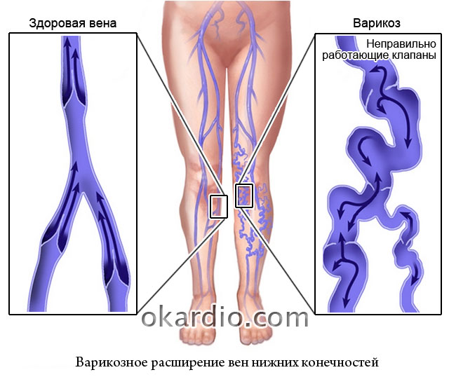
Varicose veins of superficial and deep veins begin with loss of tone and sagging of the subcutaneous vessels. This shows up:
- red “spiders” on the skin of the lower leg and thigh;
- increased fatigue of the legs;
- swelling of the feet in the evening;
- aching pain in the calves and feet.

The patient's feet at the stage of initial trophic changes
Damage to deep venous trunks leads to more pronounced symptoms:
- pain bothers me constantly;
- feet become bluish;
- thickened venous plexuses are visible under the skin.
Varicose veins are considered a female disease because they are provoked by pregnancy and increased stress on the pelvic organs, and wearing high-heeled shoes. But many men in sedentary professions (drivers, office workers) suffer from it.
A special form of varicose veins is hemorrhoids. This pathology complements bowel movement disorders due to intestinal diseases and a sedentary lifestyle. The expansion of the external hemorrhoidal veins and the internal vascular ring leads to the following consequences:
- bursting pain in the anus;
- constant itching and burning;
- bleeding during bowel movements.
Mild varicose veins are treated by:
- tonics;
- gymnastics;
- wearing compression garments;
- rubbing with ointments.
In severe cases, only surgical techniques help. The doctor selects treatment depending on the depth and diameter of the damaged vessels (sclerotherapy methods, radiofrequency ablation) or offers surgery to remove the entire vein.
Thrombophlebitis complicates the course of varicose veins by the addition of internal or external infection. The inflamed area appears red, swollen, and hot to the touch. Palpation is painful. Thrombophlebitis is most often localized in the superficial veins of the legs and arms. May be accompanied by elevated body temperature and pain.
The disease is associated with dangerous consequences - the separation of part of the blood clot, its transformation into an embolus with the flow of blood into the femoral vein, portal vein, inferior vena cava and right atrium. From here an unobstructed path to the pulmonary artery opens.
Thrombosis of the branches of the pulmonary artery leads to infarction of part of the lung, and blockage of the main vessel causes instant death. A similar complication may occur in the patient in the postoperative period. Therefore, when preparing for surgical interventions, thrombosis prevention is always carried out (tight bandaging of the legs, a course of anticoagulants).

A large non-healing ulcer on the lower leg is one of the forms of manifestation of lost nutritional functions of the skin and muscles
Diseases of the peripheral vessels of the legs in a severe stage are accompanied by gangrene of the toes and overlying parts. Therapeutic measures are designed to prevent tissue necrosis. Vascular pathology is especially severe against the background of diabetes mellitus and atherosclerosis of the femoral artery.
Is it possible to prevent vascular diseases?
To prevent damage to the vascular bed, it is necessary to maintain the tone of the walls, which means that the diet should always contain vitamins from vegetables and fruits.
Dietary requirements for the treatment of arterial atherosclerosis: exclude fatty and fried foods, sweets, and alcohol. Be sure to consume low-fat dairy products and fish.
Smoking should be treated as a serious obstacle to the treatment of the disease. Any treatment, even the most modern, will not bring success if you continue to smoke.
Movement is the key to healthy blood vessels. Daily gymnastics activates blood flow in small capillaries and peripheral veins. You should not engage in strenuous sports or prolonged physical work. Walking, swimming, Pilates are recommended.
Due to the variety of forms of diseases, vascular problems are dealt with by: cardiologists, therapists, surgeons, neurologists, phlebologists, neurosurgeons. If symptoms occur, it is necessary to undergo examination and begin treatment. This will help prevent dangerous complications.
Vascular diseases are the main topic of this article. Basic data on the occurrence, causes of occurrence, symptoms of diseases and principles of their treatment. Forecasts for various pathologies of veins and arteries.
Article publication date: 07/01/2017
Article updated date: 08/07/2019
1.
2.
3.
4.
5.
6.
7.
8.
9.
10.
11.
Pathologies of the vascular system are a large group of diseases with a prevalence ranging from 0.0014 to 30%. They have a great impact on the working population, being the main cause of premature death when cardiac or cerebral vessels are damaged.
Most vascular pathology cannot be completely cured and requires constant maintenance therapy and observation. Significantly reduces the quality of life, the ability to exercise and causes disability in patients.
Vein pathology prevails over arterial diseases, but damage to the blood-carrying vascular structures of the myocardium and brain is often fatal (more than 70%, depending on the degree of blood flow disturbance).
Not all causes of diseases are known, but a number of risk factors are common to vascular pathology:
- Family predisposition.
- Heavy weight.
- Hypertension.
- Smoking.
- Treatment with hormonal drugs.
- Diabetes.
Vascular diseases are treated by many doctors, the main specialists being: vascular and endovascular surgeons, rheumatologists, cardiologists and neurologists.
1. Acute venous thrombosis, or thrombophlebitis
The disease is associated with the formation of a clot or clot in the lumen of a vessel, which causes an inflammatory process and disruption of normal blood flow in this area.
Another name for the disease is associated with inflammatory changes - thrombophlebitis, phlebothrombosis.
| Characteristics of the pathology | Description |
|---|---|
| Occurrence | 27–30 % |
| Causes | Long period without active walking Congenital structural features of the vein wall Changes in the blood coagulation system Injuries and infections |
| Symptoms | Pain syndrome of varying severity Swelling of part of the limb Heaviness in the arm, leg Skin changes: redness, ulceration, tissue death |
| Treatment |
|
| Forecast | There is no complete cure |
2. Postthrombophlebitic syndrome
A pathological condition that develops as a complication after thrombosis and thrombophlebitis. It is associated with a violation of the structure of the walls of the veins and leads to chronic disruption of the outflow of venous blood.

3. Varicose veins of the lower extremities
A disease leading to deformation of venous vessels with a significant increase in lumen. The process progresses over time, leading to disruption of blood flow both locally and throughout the body.
| Characteristics of the pathology | Description |
|---|---|
| Occurrence | Up to 25% |
| Causes | The main reasons have not been established Only risk factors matter |
| Symptoms | Heaviness and fatigue in the legs Moderate swelling, in the first stages - non-permanent Cold skin Later, a subcutaneous network of tortuous, nodular veins appears Skin changes (inflammation, ulcers) |
| Treatment | Drug therapy:
Surgical methods of treatment:
|
| Forecast | There is no complete cure |
4. Coronary heart disease
- Nodular.
- Necrotizing.
| Characteristics of the pathology | Description |
|---|---|
| Occurrence | Up to 0.0014% in different countries |
| Causes | Damage to the vessel wall by infection or one’s own immunity for an unknown reason |
| Symptoms | High body temperature Weight loss Pain syndrome of various localizations Increased heart rate Skin rashes Cloudy, bloody urine Pain syndrome in the myocardium Disturbances of consciousness and psyche Changing sensitivity Stool disorders |
| Treatment | Drug therapy:
|
| Forecast | There is no complete cure for the disease |
In this article we will look at vascular diseases of the brain and spinal cord and their classification. The brain is the basis of the nervous system; it provides perception, transmission and processing of information, controls all functions of the body. The proper functioning of the central nervous system is determined by a complete supply of oxygen and nutrients, therefore stable blood flow is a healthy activity of all human life support systems. Pathological changes in blood circulation in the brain and spinal cord lead to serious pathologies of the entire body and increase the risk of death.
The blood flow of the central nervous system is the cerebral circulation through the vascular system.
There is a simultaneous supply of blood through four arteries - two carotid, two vertebral, which are united by anastomoses of the arterial circle. Blood coming from the carotid arteries feeds the cerebral hemispheres, blood flowing through the vertebral arteries feeds the posterior cerebral regions.
Inside, blood flow is created by the anterior, middle and posterior paired arteries of the brain; they extend from the arterial circle and regulate the blood supply to small areas. Blood supply is also provided by radial arteries and a branched network of capillaries.
Venous blood circulates through the anastomosing.
Since each element has its own function, a change in the functioning of any vessel leads to a specific disease and a separate clinical picture and its manifestations.
Classification of vascular pathologies
Classification of vascular diseases of the brain. All vascular diseases of the spinal cord and brain are divided into types according to the nature, course, and location of the disease.
The first type includes vascular diseases:
- Atherosclerotic disease;
- Hypertension;
- Hypotension;
- Immunopathological change;
- Diseases that cause circulatory problems.
The second type of disease indicates the development of pathology caused by a violation of the functional activity of blood vessels:
- Slowly progressing lesions of cerebral vessels - discirculatory type of encephalopathy;
- Damage to brain tissue as a result of high blood pressure;
- Insufficiency and abnormalities of blood flow to the brain;
Common Causes of Vascular Diseases
Each vascular disease of the spinal cord occurs due to certain reasons. Changes in the tone, patency and other functions of blood vessels lead to the development of pathology. Vascular diseases of the central nervous system have common causes:
- Wrong lifestyle;
- Low physical activity;
- Insufficiency of the cardiovascular system;
- Chronic diseases of the spine;
- Oncology;
- Congenital blood flow disorders;
- Spinal injuries;
- Traumatic brain injuries, brain contusion.

Symptoms
The first signs indicating the onset of cerebrovascular disease are so insignificant that patients attribute them to fatigue, stress, and chronic lack of sleep.
Changes in the blood vessels progress, the blood vessels of the brain become more and more distinct.
All vascular diseases have a number of common symptoms:
- Local headaches;
- Dizziness, weakness, loss of consciousness;
- High blood pressure;
- Insomnia, irritability;
- Weakening of attention and memory;
- Inadequate coordination of movements.
Diagnostic methods
An important step in determining the location, dynamics, and extent of the lesion are examinations, the results of which allow us to make a correct diagnosis and begin full-fledged treatment.
Research into cerebrovascular diseases includes:
- External examination for the presence of neurological abnormalities;
- measurement of parameters of cardiovascular activity and respiratory system;
- Ultrasound Dopplerography - study of blood flow in large and medium-sized vessels of the cerebral system;
- MR angiography - examines the state of the cerebral vascular system using a magnetic field;
- CT angiography - examines the functional state of blood circulation and anatomical changes in blood vessels;
- Electroencephalography - establishes the state of blood flow activity and transmission of nerve impulses;
- Rheoencephalography - examines the state of elasticity of vascular tissues, determines the strength of their blood flow;
- Neurosonography - examines soft tissues and brain fibers for the presence of pathologies;
- - examines the state of vascular blood flow;
- echotomography - assesses the condition of cerebral structures;
- Dopplerography - examines hemodynamic processes, determines the dynamics of vascular restoration during treatment with medications.

Common diseases
Diseases of the vascular system of the brain are pathologies that can affect a person at different ages. All types of diseases are interdependent. If they are not treated in time, they will result in a more severe disease.
Atherosclerosis
Atherosclerosis of the vascular system is a narrowing of blood vessels by atherosclerotic plaques that limit normal blood flow. It is possible that the lumen of the brain vessels may be blocked by plaques, which leads to the cessation of blood flow to part of the brain, resulting in the death of cells and tissues.
Features of the disease
- Sweating and redness of the face;
- Trimer of chin, head;
- The venous system of the fundus has a dynamic narrowing;
- Facial asymmetry;
- Unreasonable increase in blood cholesterol levels.
At the onset of atherosclerosis, the main symptom is severe pain characteristic of the entire head.
Atherosclerosis, having a sluggish course of the disease, develops into a chronic cerebrovascular accident.
Late treatment leads to complications
- impaired sensitivity and paralysis of the cerebral lobes;
- aneurysm;
- strokes.

Cerebral aneurysm is an expansion of the carotid arteries and vein of Galen as a result of the pathology of the three-layer structure of the vessel walls. It causes bleeding in the brain due to rupture of the affected vessel.
Features of symptoms
- Eye pathology (pain, strabismus, double vision, cloudiness);
- Decreased visual and auditory sensations;
- The face becomes partially numb or paralyzed;
Signs of a ruptured aneurysm
- Severe attacks of pain accompanied by nausea and vomiting;
- Hypotension;
- Increased sensitivity of vision and hearing;
- Psychomotor impairment.
Consequences
- Brain swelling;
- Hydrocephalus;
- Cerebral ischemia;
- Vascular angiospasm.
Angiospasm is a spasm of the vascular system of the brain, accompanied by their narrowing with a sharp deterioration of the condition. Characteristic features of the disease are oxygen starvation of brain cells and decreased vascular tone.
Main signs of the disease
- Headaches in the temples, forehead, and back of the head;
- Impaired consciousness;
- Feeling pain in a certain half of the body;
- Short-term amnesia.
Consequences
- Speech impairment;
- Partial amnesia;
- Disability;
- Death.

Vascular disease leading to mental disorder
Dementia is a pathology of the vascular system of the brain, leading to impaired mental state, deterioration of memory and mental abilities.
Features of the condition
- Epilepsy;
- Impaired coordination of movements;
- Deterioration of psychophysical condition.
Consequences
- Impaired speech and mental activity;
- Mental disorders;
- Injury.
Vertebral artery syndrome is a compression of the vertebrae caused by atherosclerosis and spinal injuries. Characterized by a decrease in blood supply.
Special signs
- Changes in the structure of bones and tissues of the cervical spine;
- Intervertebral hernia;
- Injuries and inflammations of the cervical spine;
- Temporary decrease in blood flow;
- Hypoplasia.
Consequences
- Ischemia;

Ischemia
Cerebral ischemia is a vascular disease characterized by a progressive deterioration of blood supply. The affected areas of the brain stop performing their function.
The main symptom of the disease is functional mental disorder.
Consequences
- Ischemic stroke;
- Brain swelling;
- Pneumonia, respiratory paralysis;
- Oligophrenia;
- Development of cardiovascular failure.
Stroke is a vascular disease characterized by a sudden disruption of blood flow to the brain. There are several types of stroke.
Ischemic stroke is caused by a lack of blood supply to an area of the brain.
Hemorrhagic stroke is a local hemorrhage caused by an aneurysm.
Subarachnoid stroke is damage to the blood circulation of the brain due to hemorrhage in the spaces of the meninges.
Consequences
- Complete or partial paralysis;
- Amnesia;
- Impaired memory, speech, vision, hearing.

Treatment methods
Treatment of cerebrovascular diseases is carried out jointly by several doctors: a neurologist, a therapist, an ophthalmologist, an ENT specialist, and a cardiologist. All treatment of neuralgia for vascular diseases of the brain and cerebral blood flow depends on the causes and symptoms that caused the pathology.
A complex consisting of medications, injections and physiotherapeutic procedures is selected.
Medicines and injections are intended to treat vascular diseases and their causes, to ensure normal blood flow and regeneration of tissues and nerve fibers. Drugs are used to relieve the symptoms of diseases. A therapeutic diet is prepared.
In particularly severe conditions, a neurosurgeon performs surgery.
After a full course of treatment. The main method of which is physiotherapeutic measures.
The best measures to prevent changes in blood vessels today are a healthy lifestyle, proper diet, sports, and annual medical examination.
Any neurological disorders require consultation and treatment prescribed by a specialist, which will ensure a quality continuation of life.
Diseases of the blood vessels of the brain are difficult to tolerate, entail irreversible processes, and lead to acute pathologies. The most important thing is the timing of the medical care provided.
Video