Autonomic nervous system. Sympathetic and Parasympathetic Nervous System The activity of the sympathetic nervous system contributes to
The sympathetic department is part of the autonomic nervous tissue, which, together with the parasympathetic, ensures the functioning of internal organs and chemical reactions responsible for the life of cells. But you should know that there is a metasympathetic nervous system, part of the autonomic structure, located on the walls of organs and capable of contracting, contacting directly with the sympathetic and parasympathetic, making adjustments to their activity.
The human internal environment is directly influenced by the sympathetic and parasympathetic nervous system.
The sympathetic division is localized in the central nervous system. Spinal nerve tissue operates under the control of nerve cells located in the brain.
All elements of the sympathetic trunk, located on two sides of the spine, are directly connected to the corresponding organs through nerve plexuses, and each has its own plexus. At the bottom of the spine, both trunks in a person are united together.
The sympathetic trunk is usually divided into sections: lumbar, sacral, cervical, thoracic.
The sympathetic nervous system is concentrated near the carotid arteries of the cervical region, in the thoracic - the cardiac and pulmonary plexus, in the abdominal cavity the solar, mesenteric, aortic, hypogastric.
These plexuses are divided into smaller ones, and from them impulses move to the internal organs.
The transition of excitation from the sympathetic nerve to the corresponding organ occurs under the influence of chemical elements - sympathins, secreted by nerve cells.
They supply the same tissues with nerves, ensuring their relationship with the central system, often having the opposite effect on these organs.
The influence that the sympathetic and parasympathetic nervous systems have can be seen from the table below:
 Together they are responsible for cardiovascular organisms, digestive organs, respiratory structures, secretions, the work of smooth muscle of hollow organs, and control metabolic processes, growth, and reproduction.
If one begins to predominate over the other, symptoms of increased excitability appear: sympathicotonia (the sympathetic part predominates), vagotonia (the parasympathetic part predominates).
Sympathicotonia manifests itself in the following symptoms: fever, tachycardia, numbness and tingling in the extremities, increased appetite without the appearance of weight loss, indifference to life, restless dreams, fear of death for no reason, irritability, absent-mindedness, decreased salivation, as well as sweating, migraine appears.
In a person, when the increased work of the parasympathetic department of the autonomic structure is activated, increased sweating appears, the skin feels cold and damp to the touch, a decrease in heart rate occurs, it becomes less than the prescribed 60 beats in 1 minute, fainting, salivation and respiratory activity increase. People become indecisive, slow, prone to depression, and intolerant.
The parasympathetic nervous system reduces the activity of the heart and tends to dilate blood vessels.
Functions
The sympathetic nervous system is a unique design of an element of the autonomic system, which, in the event of a sudden need, is capable of increasing the body’s ability to perform work functions by collecting possible resources.
As a result, the design carries out the work of organs such as the heart, reduces blood vessels, increases muscle capacity, frequency, strength of the heart rhythm, performance, and inhibits the secretory and absorption capacity of the gastrointestinal tract.

The SNS supports functions such as the normal functioning of the internal environment in an active position, coming into action during physical effort, stressful situations, illnesses, blood loss and regulates metabolism, for example, an increase in sugar, blood clotting, and others.
It is most fully activated during psychological shocks, through the production of adrenaline (enhancing the action of nerve cells) in the adrenal glands, which allows a person to react faster and more effectively to suddenly occurring factors from the outside world.
Adrenaline can also be produced when the load increases, which also helps a person cope with it better.
After coping with the situation, a person feels tired, he needs to rest, this is due to the sympathetic system, which has most fully used up the body’s capabilities, due to the increase in body functions in a sudden situation.
The parasympathetic nervous system performs the functions of self-regulation, protection of the body, and is responsible for human bowel movements.
Self-regulation of the body has a restorative effect, working in a calm state.
The parasympathetic part of the activity of the autonomic nervous system is manifested by a decrease in the strength and frequency of the heart rhythm, stimulation of the gastrointestinal tract with a decrease in glucose in the blood, etc.
By carrying out protective reflexes, it rids the human body of foreign elements (sneezing, vomiting, etc.).
The table below shows how the sympathetic and parasympathetic nervous systems act on the same elements of the body. 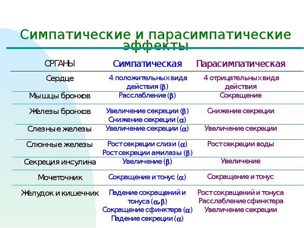
Treatment
If you notice signs of increased sensitivity, you should consult a doctor, as this can cause ulcerative, hypertensive diseases, or neurasthenia.
Only a doctor can prescribe correct and effective therapy! There is no need to experiment with the body, since the consequences if the nerves are in a state of excitability are quite a dangerous manifestation not only for you, but also for people close to you.
When prescribing treatment, it is recommended, if possible, to eliminate factors that excite the sympathetic nervous system, be it physical or emotional stress. Without this, no treatment will most likely help; after taking a course of medication, you will get sick again.
You need a cozy home environment, sympathy and help from loved ones, fresh air, good emotions.
First of all, you need to make sure that nothing raises your nerves.
The medications used in treatment primarily belong to the group of potent drugs, so they should be used carefully only as directed or after consultation with a doctor.
Prescribed medications usually include: tranquilizers (Phenazepam, Relanium and others), antipsychotics (Frenolone, Sonapax), sleeping pills, antidepressants, nootropic drugs and, if necessary, cardiac drugs (Korglikon, Digitoxin) ), vascular, sedative, vegetative drugs, a course of vitamins.
It is good to use physiotherapy, including physical therapy and massage, you can do breathing exercises and swimming. They are good at helping to relax the body.
In any case, ignoring the treatment of this disease is categorically not recommended; it is necessary to consult a doctor in a timely manner and carry out the prescribed course of therapy.
Each of us has moments of anxiety and difficult periods in life. In this book you will learn how to survive them and minimize them by “reprogramming” your brain. John Arden, a doctor with extensive experience, talks about the latest achievements and discoveries in the field of neurophysiology, describing in detail how to apply them in various areas of life to achieve success and prosperity. You'll learn healthy habits that will allow you to keep your brain active longer and lead a richer life without the limitations you impose on yourself.
This is a book for anyone who wants to learn more about their brain and improve their quality of life.
Published in Russian for the first time.
Book:
The autonomic nervous system consists of two parts: the sympathetic nervous system and the parasympathetic nervous system. The sympathetic nervous system is responsible for stimulating the body's reactions, and the parasympathetic nervous system is responsible for inhibiting the reactions. In extreme situations, the sympathetic nervous system activates the HPA axis and the fight or flight response. The action of the parasympathetic nervous system was called the relaxation response by Harvard University professor Herbert Benson. Activation of the parasympathetic nervous system leads to inhibition of cardiac activity, a slowdown in metabolic processes in the body and the level of respiration.
The active principle described earlier activates the BNST and the left frontal lobe of the prefrontal cortex. This effort creates the precondition for the parasympathetic nervous system to later ensure that the body relaxes.
The switch between the sympathetic and parasympathetic nervous systems through the prefrontal cortex and hippocampus may not occur as quickly if a person suffers from post-traumatic stress disorder (PTSD). The amygdala has heightened sensitivity to the context in which the trauma occurred. An example was previously given of a war veteran who was frightened by fireworks. But even military veterans with PTSD can tame their amygdala, as I describe in Conquering Posttraumatic Stress Disorder with Dr. Victoria Beckner.
Different types of breathing determine different emotional states. Breathing quickens when a person experiences anxiety. With a high breathing rate, the abdominal muscles tense and the sternum cavity contracts.
People sometimes come to my anti-anxiety trainings with rapid breathing. They usually tend to speak very quickly and thus prevent themselves from breathing normally. They start with some neutral topic, but soon their tone changes due to rapid breathing and a growing feeling of anxiety. Increased levels of anxiety activate memories and response patterns associated with the same networks that support anxious mental activity. Soon a new topic of conversation causes even greater anxiety.
Typically, humans have a resting respiratory rate of 9 to 16 breaths per minute. In a state of panic attack, this figure increases to 27 inhalations and exhalations per minute. As your breathing rate increases, you experience many of the symptoms associated with a panic attack, including numbness, tingling, dry mouth, and dizziness.
Since the cardiovascular system integrates the respiratory and circulatory systems, rapid breathing causes the heart rate to increase, making a person even more anxious. When your breathing slows down, your heart rate also slows down, which promotes calm and relaxation.
To learn to relax, you need to make an effort and develop some new useful habits, such as controlling your breathing. Since rapid breathing is one of the most characteristic symptoms of panic, it is worth learning how to breathe correctly. During hyperventilation, or rapid breathing, real physiological changes occur in the human body and, in particular, in the brain.
When a person hyperventilates, they inhale too much oxygen, which lowers the level of carbon dioxide in the blood. Carbon dioxide helps maintain an optimal acid-base balance (pH level) in the blood. As pH levels decrease, nerve cells become more excitable and a person may feel restless. Physical sensations, superimposed on uncontrollable anxiety, can even provoke a panic attack.
Excessive reduction in carbon dioxide levels in the blood causes a condition known as respiratory (hypocapnic) alkalosis, in which the blood is characterized by a high alkaline content and low acidity. Then a narrowing of the blood vessels occurs, as a result of which the blood supply to the organs and tissues of the body deteriorates. Hemoglobin binds oxygen tightly, resulting in tissues and organs receiving less oxygen. And here’s the paradox: despite the fact that a person inhales too much oxygen, tissues and organs receive less oxygen than needed.
Chapter 17. Antihypertensive drugs
Antihypertensives are drugs that lower blood pressure. Most often they are used for arterial hypertension, i.e. with high blood pressure. Therefore, this group of substances is also called antihypertensive drugs.
Arterial hypertension is a symptom of many diseases. There are primary arterial hypertension, or hypertension (essential hypertension), as well as secondary (symptomatic) hypertension, for example, arterial hypertension with glomerulonephritis and nephrotic syndrome (renal hypertension), with narrowing of the renal arteries (renovascular hypertension), pheochromocytoma, hyperaldosteronism, etc.
In all cases, they strive to cure the underlying disease. But even if this fails, arterial hypertension should be eliminated, since arterial hypertension contributes to the development of atherosclerosis, angina pectoris, myocardial infarction, heart failure, visual impairment, and renal dysfunction. A sharp increase in blood pressure - a hypertensive crisis can lead to bleeding in the brain (hemorrhagic stroke).
The causes of arterial hypertension are different for different diseases. In the initial stage of hypertension, arterial hypertension is associated with an increase in the tone of the sympathetic nervous system, which leads to an increase in cardiac output and constriction of blood vessels. In this case, blood pressure is effectively reduced by substances that reduce the influence of the sympathetic nervous system (central-acting antihypertensives, adrenergic blockers).
In kidney disease and in the late stages of hypertension, an increase in blood pressure is associated with activation of the renin-angiotensin system. The resulting angiotensin II constricts blood vessels, stimulates the sympathetic system, increases the release of aldosterone, which increases the reabsorption of Na + ions in the renal tubules and thus retains sodium in the body. Drugs that reduce the activity of the renin-angiotensin system should be prescribed.
With pheochromocytoma (tumor of the adrenal medulla), adrenaline and norepinephrine secreted by the tumor stimulate the heart and constrict blood vessels. Pheochromocytoma is removed surgically, but before surgery, during surgery, or if surgery is not possible, blood pressure is reduced with the help of wasp-blockers.
A common cause of arterial hypertension may be sodium retention in the body due to excessive consumption of table salt and insufficiency of natriuretic factors. An increased content of Na + in the smooth muscles of blood vessels leads to vasoconstriction (the function of the Na + /Ca 2+ exchanger is impaired: the entry of Na + and the exit of Ca 2+ decreases; the level of Ca 2+ in the cytoplasm of smooth muscles increases). As a result, blood pressure increases. Therefore, for arterial hypertension, diuretics are often used that can remove excess sodium from the body.
For arterial hypertension of any origin, myotropic vasodilators have an antihypertensive effect.
It is believed that patients with arterial hypertension should use antihypertensive drugs systematically to prevent an increase in blood pressure. For this purpose, it is advisable to prescribe long-acting antihypertensive drugs. The most commonly used drugs are those that act for 24 hours and can be prescribed once a day (atenolol, amlodipine, enalapril, losartan, moxonidine).
In practical medicine, the most commonly used antihypertensive drugs are diuretics, β-blockers, calcium channel blockers, α-blockers, ACE inhibitors, and AT 1 receptor blockers.
To relieve hypertensive crises, diazoxide, clonidine, azamethonium, labetalol, sodium nitroprusside, and nitroglycerin are administered intravenously. For mild hypertensive crises, captopril and clonidine are prescribed sublingually.
Classification of antihypertensive drugs
I. Drugs that reduce the influence of the sympathetic nervous system (neurotropic antihypertensive drugs):
1) means of central action,
2) drugs that block sympathetic innervation.
P. Vasodilators of myotropic action:
1) donors N0,
2) activators of potassium channels,
3) drugs with an unclear mechanism of action.
III. Calcium channel blockers.
IV. Agents that reduce the effects of the renin-angiotensin system:
1) drugs that interfere with the formation of angiotensin II (drugs that reduce renin secretion, ACE inhibitors, vasopeptidase inhibitors),
2) AT 1 receptor blockers.
V. Diuretics.
Drugs that reduce the influence of the sympathetic nervous system
(neurotropic antihypertensive drugs)
The higher centers of the sympathetic nervous system are located in the hypothalamus. From here, excitation is transmitted to the center of the sympathetic nervous system, located in the rostroventrolateral medulla oblongata (RVLM - rostro-ventrolateral medulla), traditionally called the vasomotor center. From this center, impulses are transmitted to the sympathetic centers of the spinal cord and further along the sympathetic innervation to the heart and blood vessels. Activation of this center leads to an increase in the frequency and strength of heart contractions (increased cardiac output) and to an increase in the tone of blood vessels - blood pressure increases.
Blood pressure can be reduced by inhibiting the centers of the sympathetic nervous system or by blocking sympathetic innervation. In accordance with this, neurotropic antihypertensive drugs are divided into central and peripheral agents.
TO centrally acting antihypertensive drugs include clonidine, moxonidine, guanfacine, methyldopa.
Clonidine (clonidine, hemitone) is an α2-adrenergic agonist, stimulates α2A-adrenergic receptors in the center of the baroreceptor reflex in the medulla oblongata (nucleus of the solitary tract). In this case, the vagal centers (nucleus ambiguus) and inhibitory neurons are excited, which have a depressing effect on the RVLM (vasomotor center). In addition, the inhibitory effect of clonidine on RVLM is due to the fact that clonidine stimulates I 1 -receptors (imidazoline receptors).
As a result, the inhibitory effect of the vagus on the heart increases and the stimulating effect of sympathetic innervation on the heart and blood vessels decreases. As a result, cardiac output and the tone of blood vessels (arterial and venous) decrease - blood pressure decreases.
Partly, the hypotensive effect of clonidine is associated with the activation of presynaptic α2-adrenergic receptors at the endings of sympathetic adrenergic fibers - the release of norepinephrine decreases.
In higher doses, clonidine stimulates extrasynaptic a 2 B -adrenergic receptors of smooth muscles of blood vessels (Fig. 45) and, with rapid intravenous administration, can cause short-term vasoconstriction and an increase in blood pressure (therefore, intravenous clonidine is administered slowly, over 5-7 minutes).
Due to the activation of α2-adrenergic receptors in the central nervous system, clonidine has a pronounced sedative effect, potentiates the effect of ethanol, and exhibits analgesic properties.
Clonidine is a highly active antihypertensive drug (therapeutic dose when administered orally 0.000075 g); lasts about 12 hours. However, when used systematically, it can cause a subjectively unpleasant sedative effect (distracted thoughts, inability to concentrate), depression, decreased tolerance to alcohol, bradycardia, dry eyes, xerostomia (dry mouth), constipation, impotence. If you abruptly stop taking the drug, a pronounced withdrawal syndrome develops: after 18-25 hours, blood pressure rises, and a hypertensive crisis is possible. β-Adrenergic blockers increase clonidine withdrawal syndrome, so these drugs are not prescribed together.
Clonidine is used mainly to quickly lower blood pressure during hypertensive crises. In this case, clonidine is administered intravenously over 5-7 minutes; with rapid administration, an increase in blood pressure is possible due to stimulation of vascular α2-adrenergic receptors.
Clonidine solutions in the form of eye drops are used in the treatment of glaucoma (reduces the production of intraocular fluid).
Moxonidine(cint) stimulates imidazoline 1 1 receptors and, to a lesser extent, a 2 adrenergic receptors in the medulla oblongata. As a result, the activity of the vasomotor center decreases, cardiac output and blood vessel tone decrease, and blood pressure decreases.
The drug is prescribed orally for the systematic treatment of arterial hypertension 1 time per day. In contrast to clonidine, moxonidine causes less pronounced sedation, dry mouth, constipation, and withdrawal symptoms.
Guanfatsin(estulik) similarly to clonidine stimulates central α2-adrenergic receptors. Unlike clonidine, it does not affect 1 1 receptors. The duration of the hypotensive effect is about 24 hours. It is prescribed orally for the systematic treatment of arterial hypertension. Withdrawal syndrome is less pronounced than with clonidine. 
Methyldopa(dopegite, aldomet) chemical structure - a-methyl-DOPA. The drug is prescribed orally. In the body, methyldopa is converted into methylnorepinephrine, and then into methyladrenaline, which stimulate the α2-adrenergic receptors of the baroreceptor reflex center.
Metabolism of methyldopa
The hypotensive effect of the drug develops after 3-4 hours and lasts about 24 hours.
Side effects of methyldopa: dizziness, sedation, depression, nasal congestion, bradycardia, dry mouth, nausea, constipation, liver dysfunction, leukopenia, thrombocytopenia. Due to the blocking effect of a-methyl-dopamine on dopaminergic transmission, the following are possible: parkinsonism, increased production of prolactin, galactorrhea, amenorrhea, impotence (prolactin inhibits the production of gonadotropic hormones). If you abruptly stop taking the drug, withdrawal symptoms appear after 48 hours.
Drugs that block peripheral sympathetic innervation.
To reduce blood pressure, sympathetic innervation can be blocked at the level of: 1) sympathetic ganglia, 2) endings of postganglionic sympathetic (adrenergic) fibers, 3) adrenergic receptors of the heart and blood vessels. Accordingly, ganglion blockers, sympatholytics, and adrenergic blockers are used.
Ganglioblockers - hexamethonium benzosulfonate(benzo-hexonium), azamethonium(pentamine), trimethaphan(arfonade) block the transmission of excitation in the sympathetic ganglia (block N N -xo-linoreceptors of ganglionic neurons), block N N -cholinergic receptors of chromaffin cells of the adrenal medulla and reduce the release of adrenaline and norepinephrine. Thus, ganglion blockers reduce the stimulatory effect of sympathetic innervation and catecholamines on the heart and blood vessels. There is a weakening of heart contractions and expansion of arterial and venous vessels - arterial and venous pressure decreases. At the same time, ganglion blockers block the parasympathetic ganglia; thus eliminating the inhibitory effect of the vagus nerves on the heart and usually causing tachycardia.
For systematic use, ganglion blockers are of little use due to side effects (severe orthostatic hypotension, impaired accommodation, dry mouth, tachycardia; possible intestinal and bladder atony, sexual dysfunction).
Hexamethonium and azamethonium act for 2.5-3 hours; administered intramuscularly or subcutaneously during hypertensive crises. Azamethonium is also administered intravenously slowly in 20 ml of isotonic sodium chloride solution for hypertensive crisis, edema of the brain, lungs against the background of high blood pressure, for spasms of peripheral vessels, for intestinal, hepatic or renal colic.
Trimetaphan acts for 10-15 minutes; administered in solutions intravenously by drip for controlled hypotension during surgical operations.
Sympatholytics- reserpine, guanethidine(octadine) reduce the release of norepinephrine from the endings of sympathetic fibers and thus reduce the stimulating effect of sympathetic innervation on the heart and blood vessels - arterial and venous pressure decreases. Reserpine reduces the content of norepinephrine, dopamine and serotonin in the central nervous system, as well as the content of adrenaline and norepinephrine in the adrenal glands. Guanethidine does not penetrate the blood-brain barrier and does not change the content of catecholamines in the adrenal glands.
Both drugs differ in their duration of action: after stopping systematic use, the hypotensive effect can last up to 2 weeks. Guanethidine is much more effective than reserpine, but is rarely used due to severe side effects.
Due to the selective blockade of sympathetic innervation, the influences of the parasympathetic nervous system predominate. Therefore, when using sympatholytics, the following are possible: bradycardia, increased secretion of HC1 (contraindicated in peptic ulcers), diarrhea. Guanethidine causes significant orthostatic hypotension (associated with a decrease in venous pressure); When using reserpine, orthostatic hypotension is mild. Reserpine reduces the level of monoamines in the central nervous system and can cause sedation and depression.
A -Adrenergic blockers reduce the stimulating effect of sympathetic innervation on blood vessels (arteries and veins). Due to the dilation of blood vessels, arterial and venous pressure decreases; heart contractions reflexively become more frequent.
a 1 -Adrenergic blockers - prazosin(minipress), doxazosin, terazosin prescribed orally for the systematic treatment of arterial hypertension. Prazosin acts for 10-12 hours, doxazosin and terazosin - 18-24 hours.
Side effects of a 1 -blockers: dizziness, nasal congestion, moderate orthostatic hypotension, tachycardia, frequent urination.
a 1 a 2 -Adrenergic blocker phentolamine used for pheochromocytoma before surgery and during surgery to remove pheochromocytoma, as well as in cases where surgery is impossible.
β -Adrenergic blockers- one of the most commonly used groups of antihypertensive drugs. When used systematically, they cause a persistent hypotensive effect, prevent sudden increases in blood pressure, practically do not cause orthostatic hypotension, and, in addition to hypotensive properties, have antianginal and antiarrhythmic properties.
β-Adrenergic blockers weaken and slow down heart contractions - systolic blood pressure decreases. At the same time, β-adrenergic blockers constrict blood vessels (block β 2 -adrenergic receptors). Therefore, with a single use of beta-blockers, the mean arterial pressure usually decreases slightly (with isolated systolic hypertension, blood pressure can decrease even after a single use of beta-blockers).
However, if p-blockers are used systematically, then after 1-2 weeks the narrowing of blood vessels is replaced by their dilation - blood pressure decreases. Vasodilation is explained by the fact that with the systematic use of beta-blockers, due to a decrease in cardiac output, the baroreceptor depressor reflex is restored, which is weakened in arterial hypertension. In addition, vasodilation is facilitated by a decrease in the secretion of renin by juxtaglomerular cells of the kidneys (block of β 1 -adrenergic receptors), as well as blockade of presynaptic β 2 -adrenergic receptors in the endings of adrenergic fibers and a decrease in the release of norepinephrine.
For the systematic treatment of arterial hypertension, long-acting β 1-blockers are often used - atenolol(tenormin; lasts about 24 hours), betaxolol(valid up to 36 hours).
Side effects of β-blockers: bradycardia, heart failure, difficulty in atrioventricular conduction, decreased HDL levels in the blood plasma, increased bronchial and peripheral vascular tone (less pronounced with β 1 -blockers), increased effect of hypoglycemic agents, decreased physical activity.
a 2 β -Adrenergic blockers - labetalol(trandate), carvedilol(Dilatrend) reduce cardiac output (block of β-adrenoreceptors) and reduce the tone of peripheral vessels (block of α-adrenoreceptors). The drugs are used orally for the systematic treatment of arterial hypertension. Labetalol is also administered intravenously during hypertensive crises.
Carvedilol is also used for chronic heart failure.
© R.R. Wenzel, Yu.V. Furmenkova, 2002
UDC 611.839-08
Received November 8, 2001
R.R. Wenzel, Yu.V. Furmenkova
State Medical Academy, Nizhny Novgorod;
University Hospital, Essen (Germany)
Antihypertensive drugs and the sympathetic nervous system
The sympathetic nervous system (SNS) is an important regulator of cardiovascular activity. Its activity is determined by psychological, nervous and humoral factors. Activation of neurohumoral systems, as well as disruption of local regulatory mechanisms, plays an important role in the development and prognosis of cardiovascular diseases.
SNS activity increases with age, regardless of the presence of pathological conditions 2 . In congestive heart failure, significant increases in sympathetic activity correlate with mortality rates 3 . Hypersympathicotonia contributes to the development of myocardial ischemia due to reflex tachycardia and narrowing of the coronary vessels, combined with the presence of arterial hypertension (AH), insulin resistance and a high risk of developing cardiovascular complications 4, 5. Although the contribution of the SNS to the development of hypertension is controversial, the role of hypersympathicotonia in the early stages of the disease is beyond doubt 6-8. It is believed that essential hypertension is associated with increased sympathetic activity at the level of the central nervous system 2, 7, 9. However, it is possible that as a result of the interaction of neuronal plexuses and pathways involved in the regulation of sympathetic activity at the central level, blood pressure (BP) and the risk of vascular complications may be reduced. Pharmacotherapy of hypertension and its effect on the activity of the SNS served as the topic of this article.
Regulation of the sympathetic nervous system
Efferent fibers of the medulla oblongata connect it to the vasomotor center. Innervation of internal organs is carried out by two neurons united in a ganglion. Myelinated axons of preganglionic neurons of the thoracic and lumbar spinal cord approach postganglionic neurons of the sympathetic trunk and prevertebral ganglia. The mediator of nerve impulse transmission from the presynaptic to the postsynaptic neuron is acetylcholine, which binds to nicotine-sensitive receptors. The mediator of adrenergic receptors, norepinephrine, participates in the transmission of impulses to effector organs.
The catecholamines epinephrine, norepinephrine and dopamine are produced in the adrenal glands, which are phylogenetically a ganglion. In peripheral vessels, sympathetic activation causes vasoconstriction, mediated by the action of beta-adrenergic receptors on smooth muscle cells and beta-adrenergic receptors on the heart. Experimental and early clinical data have shown that a2-adrenergic receptors have a secondary role in the sympathetic regulation of the cardiovascular system, but endothelial a2-adrenergic receptors are directly involved in adrenergic vasoconstriction 10, 11.
The SNS interacts with the renin-angiotensin system (RAS) and the vascular endothelium. Angiotensin (AT) II influences the release and reuptake of norepinephrine by presynaptic receptors 12 and activates the SNS through central mechanisms 13 , 14 . Moreover, stimulation of b1-adrenergic receptors of the juxtaglomerular apparatus leads to activation of the RAS due to an increase in renin concentration 15 ; this mechanism, as well as sodium and water retention, contributes to an increase in blood pressure.
In addition to histamine, dopamine and prostaglandins, the production of norepinephrine in presynaptic receptors is inhibited by norepinephrine itself through a feedback regulation mechanism, while the presynaptic release of norepinephrine is stimulated by epinephrine and AT II.
Methods for studying the activity of the sympathetic nervous system
There are various ways to study the activity of the SNS. Well-known indirect methods include measurements of blood pressure, blood flow velocity and heart rate (HR). However, the interpretation of these data is difficult, since the reaction of effector organs to changes in sympathetic activity is slow and also depends on local chemical, mechanical and hormonal influences. In clinical practice, SNS activity is determined by the concentration of norepinephrine in the blood plasma. But the level of norepinephrine as an adrenergic neurotransmitter released from synaptic endings is also an indirect indicator. In addition, plasma concentrations of norepinephrine reflect the activity of not only adrenergic neurons, but also the adrenal glands. Methods for measuring plasma catecholamines have varying degrees of accuracy 16 , so other methods such as heart rate variability and blood pressure studies are worth considering 17 , 18 .
Microneurography allows direct determination of cutaneous or muscular sympathetic activity in the peripheral nerve 19 , 20 . Nerve impulses are recorded at the moment of their occurrence, and it is possible not only to observe their changes in response to stimulation, but also to monitor them 19-23. This is a direct method of measuring SNS activity in the medulla oblongata. New advances in microneurography make it possible to characterize changes in the activity of sympathetic nerves in response to cardiovascular drugs and analyze the pharmacokinetic capabilities of the latter 24 .
In addition, information about the influence of the SNS on effector organs is provided by measuring systolic intervals, cardiac impedanceography, plethysmography and laser Dopplerography 16, 25-28.
Effect of drugs on the sympathetic nervous system
Beta blockers
β-Adrenergic receptor antagonists reduce the positive inotropic and chronotropic effects of catecholamines mediated through β1-adrenergic receptors and β2-adrenergic relaxation of vascular smooth muscle cells 29-32. In addition, blockade of b-adrenergic receptors inhibits the metabolic effects of catecholamines such as lipolysis or glycogenolysis 31.
In the treatment of cardiovascular diseases, selective blockade of b1 receptors protects the heart from excessive sympathetic stimulation, reducing the frequency and force of heart contractions, and as a result, myocardial oxygen consumption 31.
Beta-blockers are the drugs of choice in the treatment of hypertension and coronary heart disease (CHD) because they reduce mortality, the incidence of ischemic episodes, the risk of primary and recurrent myocardial infarction, and sudden coronary death 33-36.
In recent years, β-adrenergic antagonists have been used in the treatment of congestive heart failure 37–39. The positive effect of b-adrenergic receptor blockade in heart failure, leading, apparently, to better functioning of the SNS, is observed with bisoprolol 40, metoprolol 41 and carvedilol 42. It has been proven that these drugs not only improve hemodynamics and clinical symptoms, but also reduce mortality 42, 43, although at the beginning of treatment, during the selection of an adequate dose in cases of severe heart failure, mortality may increase. Thus, β-adrenergic receptor antagonists improve the sensitivity of the latter to their agonists 44. On the central link of the sympathetic nervous system, b-blockade has the opposite effect, which has not been studied enough 45, 46. Although sympathetic nerve activity increased with intravenous administration of the β1-selective β-blocker metoprolol to patients with untreated hypertension 45 , it decreased with long-term use of this drug 46 . Interestingly, the effect of selective b1- and non-selective b-blockers on SNS activity differs, at least after the first dose in healthy volunteers. At the same time, the level of catecholamines in plasma increases significantly after administration of the beta-selective beta-blocker bisoprolol, while administration of the non-selective beta-blocker propranolol does not affect the plasma concentration of norepinephrine 29, 31.
Diuretics
Diuretics inhibit the reabsorption of salts and water in the tubules, which reduces pre- and afterload. The increased release of salt and water ions under the influence of diuretics activates not only vasopressin, the renin-angiotensin-aldosterone system, but also the SNS, which compensates for disturbances in the water-salt balance 47.
Nitrates
Nitrates, as peripheral vasodilators, cause endothelium-dependent relaxation of vascular smooth muscle cells. Side effects of some drugs in this group include reflex tachycardia. In a double-blind, placebo-controlled study, isosorbide dinitrate markedly increased both heart rate and, as measured by microneurography, SNS activity 24 . This confirms the results of studies of the effects of other vasodilators when administered intravenously 48-50. This effect can be explained by the fact that, following a possible decrease in central venous pressure, pulse pressure decreases and baroreceptors are activated 24 .
Other vasodilators, including a1-blockers
The vasodilators minoxidil and hydrolasine effectively lower blood pressure by reducing pre- and afterload. However, they stimulate the SNS, so during long-term treatment, compensatory activation of the sympathetic and renin-angiotensin systems predominates 51 .
Selective α1-adrenergic receptor antagonists, such as prazosin, also reduce pre- and afterload by inhibiting peripheral sympathetic vasoconstriction, but do not affect the sympathetic activity of the myocardium, since it contains mainly β-adrenergic receptors 52. This explains why the Veterans Administration Cooperative Study (VACS) trial, which used prazosin, did not demonstrate an improvement in prognosis in patients with heart failure 53 . It should be noted that the α1-adrenergic receptor antagonist doxazosin significantly activates the SNS, both at rest and during exercise, compared to placebo 29, 54.
Calcium ion antagonists
Calcium antagonists (CAs) cause peripheral vasodilation and inhibition of the effect of vasoconstrictors on smooth muscle due to blockade of slow L-type calcium channels and a decrease in calcium ion transport. A decrease in the intracellular concentration of the latter inhibits electromechanical processes, which leads to vasodilation and a decrease in blood pressure. Representatives of three groups of calcium antagonists - dihydropyridine (nifedipine), phenylalkylamine (verapamil) and benzodiazipine (diltiazem) types bind different parts of the α1 subunit of the calcium channel. If drugs of the dihydropyridine group are predominantly peripheral vasodilators, then substances like verapamil may directly act on the sinoatrial node and probably reduce the activity of the SNS.
AA have positive antihypertensive and antiischemic effects 55 . Moreover, they have vasoprotective capabilities, improve endothelial function in atherosclerosis and hypertension, both experimentally and in the treatment of patients with hypertension 56, 57. AA inhibit the proliferation of smooth muscle cells in human coronary arteries 58 and, to some extent, the progression of atherosclerosis 59–67.
Despite the vasoprotective effect, clinical studies of AK in patients with coronary artery disease, impaired left ventricular function, and diabetes did not give a positive result 60-67.
Activation of the SNS depends not only on the group of AAs used, but also on their pharmacokinetics. For example, dihydropyridine AKs (i.e. nifedipine, felodipine, amlodipine) increase SNS activity and cause reflex tachycardia 68, 69. On the contrary, verapamil reduces heart rate and, as shown by plasma norepinephrine studies, SNS activity 70 . A single dose of nifedipine to healthy volunteers, according to microneurography, increased the tone of the SNS, which was typical for both short- and long-acting drugs. However, nifedipine has different effects on the sympathetic nerves leading to the heart and blood vessels. Thus, heart rhythm was not an accurate indicator of the state of the nervous system and a slight increase in heart rate did not indicate a decrease in sympathetic activity 68 .
Amlodipine, a new long-acting AA, appears to stimulate the SNS to a lesser extent than other dihydropyridine drugs. Although heart rate and plasma norepinephrine levels in hypertensive patients increased significantly during an acute drug test with amlodipine, no effect on heart rate was observed with long-term use 69 .
Angiotensin-converting enzyme inhibitors
By blocking the enzyme, angiotensin-converting enzyme (ACE) inhibitors disrupt the synthesis of AT II, a powerful vasoconstrictor that increases the release of norepinephrine by stimulating peripheral presynaptic receptors 71. Moreover, AT II stimulates the activity of the central division of the SNS 72 . It is believed that ACE inhibitors also prevent inhibition of bradykinin synthesis and thereby promote vasodilation. Bradykinin promotes the release of nitric oxide and prostacyclin from the endothelium, which enhances the hemodynamic response to ACE blockade. However, bradykinin can also have side effects, in particular cough and vascular edema 73-77.
Unlike vasodilators (nitrates or calcium antagonists) that activate the SNS, ACE inhibitors do not cause reflex tachycardia and increase plasma norepinephrine levels 78 . In a double-blind, placebo-controlled study, the ACE inhibitor captopril, administered intravenously to healthy volunteers, reduced sympathetic nerve activity despite lowering blood pressure and did not alter the response to mental or physical stress, whereas nitrates caused significant activation of the SNS 3, 24. Thus, a decrease in the plasma concentration of AT II, which stimulates the activity of the SNS, reduces the tone of the SNS 72. This is the only possible explanation for the beneficial effect of ACE inhibitors on survival in patients with left ventricular dysfunction, in whom increased SNS tone was associated with high mortality 79 . The beneficial effects of ACE inhibitors on morbidity and mortality in patients with heart failure and left ventricular dysfunction, as well as in patients with myocardial infarction, have been reported in many clinical studies 79–83.
However, there are a number of mechanisms that partially offset the beneficial effects of ACE inhibitors noted with acute intravenous administration. First of all, AT II can be synthesized in an alternative way, independent of ACE, with the help of chymases; at the same time, the SNS is inhibited to a lesser extent 84-86. On the other hand, it has been established that chronic ACE inhibition does not alter the biosynthesis, accumulation and release of catecholamines 87. Since bradykinin dose-dependently stimulates the release of norepinephrine, even during blockade of the converting enzyme, it can be considered to compensate for the lack of effect of ACE inhibitors by promoting the release of catecholamines 87. In heart failure, chronic treatment with ACE inhibitors is accompanied by a marked decrease in central sympathetic activity, possibly due to the effect of constantly stressed baroreflex mechanisms on the SNS 88 . The activity of the parasympathetic nervous system does not appear to change with acute and chronic administration of ACE inhibitors, since these drugs do not affect basic cardiovascular reflexes 89 .
Angiotensin type I receptor antagonists
Blockade of AT II receptors is the most direct way to inhibit the RAS. Unlike ACE inhibitors, which do not affect the release of norepinephrine due to inhibition of its reuptake and metabolism, activation of compensatory mechanisms, angiotensin type I receptor antagonists (ATI) in vitro suppress angiotensin-induced norepinephrine uptake and, therefore, its proliferative effect 90, 91.
The effect of AT I receptor antagonists in the human body in vivo has not yet been sufficiently studied. A study of the efficacy of losartan in the elderly showed that the AT I receptor antagonist losartan had a greater effect on morbidity and mortality in patients with symptomatic heart failure than the ACE inhibitor captopril 92 . There were no differences in plasma concentrations of norepinephrine between the groups of patients receiving losartan and captopril.
Experimental data have shown that AT I receptor antagonists suppress catecholamine synthesis to a greater extent than ACE inhibitors 93 . It has been established that the new non-peptide AT I receptor antagonist eprosartan inhibits the pressor response to spinal cord stimulation in rats, while losartan, valsartan and irbesartan do not affect the SNS. This fact can be regarded as a more pronounced inhibition of AT II receptors 94 .
It is unknown whether these effects on the SNS will be significant in vivo. However, the first clinical results of a double-blind, placebo-controlled study showed that, at least, losartan did not reduce SNS activity at rest or after exercise compared with placebo or enalapril 54 .
Central sympatholytics
Clonidine, guafacin, guanabenz and a-methyl-DOPA are well-known antihypertensive drugs that act on central α2-adrenergic receptors 95 and lead to depression of the SNS and a decrease in blood pressure, mainly as a result of vasodilation and a subsequent decrease in peripheral vascular resistance. Despite their good hypotensive effect, these substances are no longer used as first-line agents in the treatment of hypertension due to their unwanted side effects such as nausea, dry mouth and drowsiness. Withdrawal syndrome is also possible with clonidine use 96 . These side effects are mainly related to the action on α2-adrenergic receptors 97 .
Clinical use of a new generation of centrally acting antihypertensive drugs (for example, moxonidine and rilmenidine) with fewer side effects has now begun. It has been established that they have a greater effect on central imidazoline1 receptors than on a2-adrenergic receptors 97-99. In contrast, other centrally acting antihypertensive drugs (α-methyl-DOPA, guanfacine, guanabenz) interact predominantly with central α2 receptors 95 . In laboratory animals, moxonidine inhibited the sympathetic innervation of resistive vessels, the heart and kidneys 97, 100. A double-blind, placebo-controlled in vivo study with direct measurement of SNS activity using microneurography demonstrated for the first time that the imidazoline-1 receptor agonist moxonidine reduces systolic and diastolic blood pressure due to a decrease in central SNS tone in both healthy volunteers and untreated hypertensive patients 68 . Moxonidine reduces sympathetic activity and plasma norepinephrine levels in both groups, while the concentrations of epinephrine and renin did not change 68. Heart rate after taking moxonidine decreased in healthy individuals; in patients with hypertension, a tendency to bradycardia was observed only at night 68.
In terms of its ability to control blood pressure, moxonidine is comparable to other antihypertensive drugs, such as a- and b-blockers, calcium antagonists or ACE inhibitors; side effects (nausea, dry mouth) are less pronounced than with clonidine and other centrally acting drugs of the previous generation 30, 101.
Rilmenidine is another imidazoline 1 receptor agonist with even greater affinity for the latter 102 . Its use in patients has shown effective blood pressure lowering with fewer side effects than clonidine 103-105. Rilmenidine caused the same reduction in blood pressure as the beta-adrenergic receptor antagonist atenolol, but was better tolerated by patients compared to it. However, unlike atenolol, it did not affect measures of autonomic nervous system function such as heart rate during exercise and the Valsalva maneuver 106 . The effect of rilmenidine on the central nervous system has not yet been studied.
Interaction between the sympathetic nervous system and the vascular endothelium
The vascular endothelium plays an important role in regulating their tone. Impaired secretion of mediators by the endothelium may be one of the links in the pathogenesis and progression of hypertension and atherosclerosis. Experimental data have shown the presence of a variety of interactions between the SNS and the vascular endothelium. Endothelin-1, produced by endothelial cells, is a powerful vasoconstrictor; its plasma concentration correlates with mortality rates from severe cardiovascular disease 107 , 108 . Endothelin causes peripheral vasoconstriction and increased blood pressure; in rats, endothelin administration stimulates sympathetic activity 109 . In addition, this substance is considered a comitogen for the proliferation of vascular smooth muscle cells 108.
Endothelin receptors are coupled to calcium channels via G proteins 110 . This fact may explain how calcium antagonists reduce endothelium-dependent vasoconstriction. A blood flow study in the forearm showed that verapamil or nifedipine administered intraarterially prevented the constrictor response to intravenous endothelin infusion 28 . On the other hand, drugs that activate the SNS (eg, nitrates and nifedipine) increase plasma endothelin concentrations in humans, whereas ACE inhibitors and moxonidine inhibit SNS activity and do not affect endothelin levels 24, 111.
Long-term therapy with calcium antagonists experimentally and in patients with hypertension improves endothelium-dependent relaxation in response to acetylcholine 112 . ACE inhibitors also stimulate endothelium-dependent relaxation by inhibiting the inactivation of bradykinin, which leads to the formation of nitric oxide and prostacyclin. When studying blood flow in resistive vessels in rats with spontaneous hypertension, it was found that long-term blockade of the RAS with the non-peptide AT II receptor antagonist CGP 48369, the ACE inhibitor benazepril or the calcium antagonist nifedipine reduced blood pressure and improved endothelial function 56 . Clinical studies have shown that the ACE inhibitor quinapril is able to reverse diastolic dysfunction and reduce the incidence of coronary ischemia 113–115. Administration of the ACE inhibitor lisinopril to patients with essential hypertension selectively enhances vasodilation in response to bradykinin 116 .
Different ACE inhibitors, such as quinapril and enalapril, improve endothelium-dependent vasodilation to varying degrees, apparently having different affinities for ACE. This is supported by the fact that quinapril, in contrast to enalapril, promotes vascular dilation in patients with chronic heart failure by increasing nitric oxide 117 .
Experimental and early clinical studies of the cutaneous microcirculation in humans suggest that adrenergic agonists stimulate endothelial α-receptors and this leads to the release of nitric oxide 10, 118. Indeed, a1 receptor-mediated constriction of vascular smooth muscle cells is enhanced by nitric oxide inhibition both in vitro and in vivo 10, 118. This mechanism may have pathophysiological significance in the development of atherosclerosis and hypertension when endothelial function is impaired. The effect of other drugs on the endothelium has not yet been clarified.
Conclusion
The effects of cardiovascular drugs on the SNS are important. However, in most cases, SNS activity has been studied using indirect methods, such as analysis of heart rate variability or plasma catecholamines. In contrast, microneurography allows direct assessment of the conduction of nerve impulses along central sympathetic fibers.
The complex effect of antihypertensive drugs on pressor systems (SNS, RAS and endothelin) is clinically important, especially in the treatment of patients with diseases of the cardiovascular system. Activation of the SNS is a possible cause of side effects of many drugs. The fact that plasma norepinephrine levels predict death in patients with heart failure 3, 119, 120 suggests that they have increased SNS activity, which is also possible in other patients, especially those with hypertension 121. In addition, SNS hyperactivity can be detected in patients with diabetes mellitus and coronary artery disease, including acute coronary syndrome 122 .
The answer to the question whether the positive effect of antihypertensive drugs on the sympathetic nervous system reduces cardiovascular and overall mortality can be obtained through invasive studies.
Literature
Converse R.J., Jacobsen T.N., Toto R.D. et al. Sympathetic overactivity in patients with chronic renal failure. N Engl J Med 1992; 327: 1908-1912.
Yamada Y., Miyajima E., Tochikubo O., Matsukawa T. et al. Age-related changes in muscle sympathetic nerve activity in essential hypertension. Hypertens 1989; 13: 870-877.
Cohn J.N., Levine T.B., Olivari M.T. et al. Plasma norepinephrine as a guide to prognosis in patients with chronic congestive heart failure. N Engl J Med 1984; 311:819-823.
Neri Serneri G.G., Boddi M., Arata L. et al. Silent ischemia in unstable angina is related to an altered cardiac norepinephrine handling. Circulation 1993; 87: 1928-1937.
Julius S., Gudbrandsson T. Early association of sympathetic overactivity, hypertension, insulin resistance, and coronary risk. J Cardiovascular Pharmacol 1992; 20(Suppl 8): 40-48.
Noll G., Wenzel R.R., Schneider M. et al. Increased activation of sympathetic nervous system and endothelin by mental stress in normotensive offspring of hypertensive parents. Circulation 1996; 93:866-869.
Anderson E.A., Sinkey C.A., Lawton W.J., Mark A.L. Elevated sympathetic nerve activity in bordeline hypertensive humans. Evidence from direct intraneural recordings. Hypertens 1989; 14: 177-183.
Philipp T., Distler A., Cordes U. Sympathetic nervous system and blood pressure control in essential hypertension. Lancet 1978; 11: 959-963.
Wallin B.G., Morlin C., Hjemdahl P. Muscle sympathetic activity and venous plasma noradrenaline concentration during static exercise in normotensive and hypertensive subjects. Acta Physiol Scand 1987; 129: 489-497.
Wenzel R.R., Bruck H., Schaefers R.F., Michel M.C. The nitric-oxide inhibitor L-NMMA potenciates norepinephrine-induced vasoconstriction: effects of the alpha2-blocker yohimbine. Kidney Blood Press Res 1998; 21: 336-398.
Chen H.I., Li H.A.T., Chen C.C. Physical conditioning decreases norepinephrine-induced vasoconstriction in rabbits. Possible roles of norepinephrine-evoked endothelium-derived relaxing factor. Circulation 1994; 90:970-975.
Hilgers K.F., Veelken R., Rupprecht G., Reeh P.W. et al. Angiotensin II facilitates sympathetic transmission in rat hind limb circulation. Hypertens 1993; 21: 322-328.
Kannan H., Nakamura T., Jin X.J., Hayashida Y. et al. Effects of centrally administered angiotensin on sympathetic nerve activity and blood flow to the kidney in conscious rats. J Auton Nerv Syst 1991; 34: 201-210.
Davis J.O., Freeman R.H. Mechanisms regulating renin release. Physiol Rev 1976; 56: 1-56.
Weber F., Brodde O.E., Anlauf M., Bock K.D. Subclassification of human beta-adrenergic receptors mediating renin-release. Clin Exp Hypertens 1983; 5: 225-238.
Schaefers R.F., Nuernberger J., Wenzel R.R., Philipp T. Characterization of adrenoreceptors mediating cardiovascular and in vivo effects of a-methylnoradrenaline (AMN) in humans. Naunun-Schmiedelberg's Arch Pharmacol 1997; 356:52.
Pagani M., Lombardi F., Guzzetti S. et al. Power spectral analysis of heart rate and arterial pressure variabilities as a marker of sympathovagal interaction in man and conscious dogs. Circ Res 1986; 59: 178-193.
Esler M., Jennings G., Korner P., Blombery P. et al. Measurement of total and organ-specific norepinephrine kinetics in humans. Am J Physiol 1984; 247: 21-28.
Delius W., Hagbarth K.E., Hongell A., Wallin B.G. Manoeuvres affecting sympathetic outflow in human skin nerves. Acta Physiol Scand 1972; 84: 177-186.
Delius W., Hagbarth K.E., Hongell A., Wallin B.G. General characteristics of sympathetic activity in human muscle nerves. Acta Physiol Scand 1972; 84: 65-81.
Wallin B.G. Intraneural recordings of normal and abnormal sympathetic activity in man. In: S.R. Bannister, edit. Autonomous failure. Oxford University Press; 1988; 177-195.
Victor R.G., Leimbach W.J., Seals D.R., Wallin B.G. et al. Effects of the cold pressor test on muscle sympathetic nerve activity in humans. Hypertens 1987; 9: 429-436.
Mark A.L., Victor R.G., Nerhed C., Wallin B.G. Microneurographic studies of the mechanisms of sympathetic nerve responses to static exercise in humans. Circ Res 1985; 57: 461-469.
Noll G., Wenzel R.R., de Marchi S., Shaw S. et al. Differential effects of captopril and nitrates on muscle sympathetic nerve activity in healthy volunteers. Circulation 1997; 95: 2286-2292.
Li Q., Belz G.G. Systolic time intervals in clinical pharmacology. Eur J Clin Pharmacol 1993; 44: 415-421.
Wenzel R.R., Duthiers N., Noll G., Bucher J. et al. Endothelin and calcium antagonists in the skin microcirculation of patients with coronary artery disease. Circulation 1996; 94: 316-322.
Creager M.A., Cooke J.P., Mendelsohn M.E. et al. Impaired vasodilation of forearm resistance vessels in hypercholesterolemic humans. J Clin Invest 1990; 86: 228-234.
Kiowski W., Luescher T.F., Linder L., Buehler F.R. Endothelin-1-induced vasoconstriction in humans. Reversal by calcium channel blockade but not by nitrovasodilators or endothelium-derived relaxing factor. Circulation 1991; 83: 469-475.
Schaefers R.F., Poller U., Ponicke K. et al. Influence of adrenoceptor and muscarinic receptor blockade on the cardiovascular effects of exogenous noradrenaline and of endogenous noradrenaline released by infused tyramine. Naunyn Schmiedeberg's Arch Pharmacol 1997; 355: 239-249.
Schaefers R.F., Loew-Kroeger A., Philipp T. Wirksamkeit und vertraeglichkeit des neuen zentralwirksamen antihypertensivums moxonidin im vergleich zu enalapril. Nieren Hochdruck 1994; 23: 221-224.
Schaefers R.F., Nuernberger J., Herrmann B., Wenzel R.R. et al. Adrenoceptors mediating the cardiovascular and metabolic effects of alpha-methylnoradrenaline in man. J Pharmacol Exp Ther 1999; 289:918-925.
Schaefers R.F., Adler S., Dail A. et al. Positive inotropic effects of the beta-2-adrenoceptor antagonist treatment. J Am Coll Cardiol 1994; 23: 1224-1233.
ISIS-1. Randomized trial of intravenous atenolol among 16027 cases of suspected acute myocardial infarction: ISIS-1. First International Study of Infarct Survival Collaborative Group. Lancet 1986; 17: 57-66.
Wikstrand J., Warnold I., Olsson G., Tuomilehto J. et al. Primary prevention with metoprolol in patients with hypertension. Mortality results from the MAPHY study; JAMA 1988; 259: 1976-1982.
The IPPSH Collaborative Group I. Cardiovascular risk and risk factors in a randomized trial of treatment based on the beta-blocker oxprenolol: the International Prospective Primary Prevention Study in Hypertension (IPPSH). The IPPSH Collaborative Group. J Hypertens 1985; 3: 379-392.
Erne P., Zuber M., Schuepfer G. Betablocker und koronare Herzkrankheit. In: T. F. Luescher, ed. It. Praeventive Kardiologie in Klinik und Praxis. Bern: Verlag Hans Huber; 1993: 231-234.
Waagstein F., Hjalmarson A., Varnauskas E., Wallentin I. Effect of chronic beta-adrenergic receptor blockade in congestive cardiomyopathy. Br Heart J 1975; 37: 1022-1036.
Engelmeier R. S., O, Connel J. B., Wals R., Rad N. et al. Improvement in symptoms and exercise tolerance by metoprolol in patients with dilated cardiomyopathy. A double-blind, randomized, placebo-controlled trial. Circulation 1985; 72:536-546.
Gilbert E.M., Anderson J.L., Deitchman D. et al. Long-term beta-blocker vasodilator therapy improves cardial function in idiopathic dilated cardiomyopathy. A double-blind, randomized study of bucindolol versus placebo. Am J Med 1990; 88: 223-229.
CIBIS Investigators and Committees. A randomized trial of beta-blockade in heart failure. The cardiac insufficiency bisoprolol study (CIBIS). Circulation 1994; 90: 2153-2156.
Waagstein F., Bristow M.R., Swedberg K. et al. for the metoprolol in dilated cardiomyopathy (MDC) trial study group. Beneficial effects of metoprolol in idiopathic dilated cardiomyopathy. Lancet 1993; 342: 1441-1446.
Packer M., Bristow M.R., Cohn J.N. et al. for the U.S. Carvedilol Heart Failure Study Group. The effect of carvedilol on morbidity and mortality in patients with chronic heart failure. N Engl J Med 1993; 334: 1349-1355.
Lechat P., Escolano S., Goldmard J.L. et al. Prognostic value of bisoprolol-induced hemodynamic effects in heart failure during the Cardiac Insufficiency-Bisoprolol Study (CIBIS). Circulation1997; 96:2197-2205.
Heilbrunn S.M., Shah P., Bristow M.R., Valantine H.A. et al. Increased beta-receptor density and improved hemodynamic response to catecholamine stimulation during long-term metoprolol therapy in heart failure from dilated cardiomyopathy. Circulation 1989; 79: 483-490.
Sundlof G., Wallin B.G., Stromgren E., Nerhed C. Acute effects of metoprolol on muscle sympathetic activity in hypertensive humans. Hypertens 1983; 5: 749-756.
Wallin B.G., Sundlof G., Stromgren E., Aberg H. Sympathetic outflow to muscles during treatment of hypertension with metoprolol. Hypertens 1984; 6:557-562.
Burnier M., Brunner H.R. Neurohormonal consequences of diuretics in different cardiovascular syndromes. Eur Heart J 1992; 13(Suppl G): 28-33.
Sanders J.S., Ferguson D.W. Diastolic pressure determines autonomic response to pressure perturbation in humans. J Appl Physiol 1989; 66:800-807.
Ferguson D.W., Hayes D.W. Nifedipine potentiates cardiopulmonary baroreflex control of sympathetic nerve activity in healthy humans. Circulation 1989; 80; 285-298.
Hoffman R.P., Sinkey C.A., Kienzle M.G., Anderson E.A. Muscle sympathetic nerve activity is reduced in IDDM before overt autonomic neuropathy. Diabetes 1993; 42; 375-380.
Packer M. Vasodilatator and inotropic drugs for the treatment of chronic congestive heart failure – distinguishing hype from hope. J Am Coll Cardiol 1988; 12: 1299-1317.
Mettauer B., Rouleau J.L., Bichet D. et al. Differential long-term intrarenal and neurohumoral effects of captopril and prazozin in patients with chronic congestive heart failure – importance of initial plasma renin activity. Circulation 1986; 73: 492-502.
Cohn J.N., Archibald D.G., Ziesche S. et al. Effect of vasodilator therapy on mortality in chronic congestive heart failure. Results of a Veterans Administration Cooperative Study. N Engl J Med 1986; 314:1547-1552.
Wenzel R.R., Wambach C., Schaefers R.F. et al. Doxasosin, but not losartan or enalapril, increases exercise-induced sympathetic activation. Kidney Blood Press Res 1998; 21: 336-398.
Nayler W.G., Szeto J. Effect of verapamil on contractility, oxygen utilization and calcium exchangeability in mammalian heart muscle. Cardiovasc Res 1972; 6: 120-128.
Dohi Y., Criscione L., Pfeiffer K., Luescher T.F. Angiotensin blockade or calcium antagonists improve endothelial dysfunction in hypertension: studies in perfused mesenteric resistance arteries. J Cardiovasc Pharmacol 1994; 24: 372-379.
Taddei S., Virdis A., Ghiadoni L., Salvetti A. Endothelial dysfunction in hypertension: fact or fancy?
J Cardiovasc Pharmacol 1998; 32 (suppl 3): 41-47.
Yang Z., Noll G., Luescher T.F. Calcium antagonists inhibit proliferation of human coronary smooth muscle cells in response to pulsatile stretch and platelet-derived growth factor. Circulation 1993; 88: 832-836.
Lichten P.R., Hugenholtz P.C., Rafflenbeul W., Hecker H. et al. Retardation of angiographic progression of coronary artery disease by nifedipine. Results of the International Nifedipine Trial on Antiatherosclerotic Therapy (INTACT). INTACT Group Investigators. Lancet 1990; 335: 1109-1113.
HINT. Early treatment of unstable angina in the coronary care unit: a randomized, double blind, placebo controlled comparison of recurrent ischaemia in patients treated with nifedipine or metoprolol or both. Report of The Holland Interuniversity Nifedipine/Metoprolol Trial (HINT) Research group. Br Heart J 1986; 56: 400-413.
Behar S., Rabinowitz B., Zion M. et al. Immediate and long-term prognostic significance of a first anterior versus first inferior wall Q-wave acute myocardial infarction. Secondary Prevention Reinfarction Israeli Nifedipine Trial (SPRINT) Study Group. Am J Cardiol 1993; 72; 1366-1370.
Estacio R.O., Schrier R.W. Antihypertensive therapy in type 2 diabetes: implications of the appropriate blood pressure control in diabetes (ABCD) trial. Am J Cardiol 1998; 82:9-14.
SPRINT. Secondaryprevention reinfarction Israeli nifedipine trial (SPRINT). A randomized interventional trial of nifedipine in patients with acute myocardial infarction. The Israeli Sprint Study Group. Eur Heart J 1988; 9: 354-364.
Tatti P., Pahor M., Byington R.P. et al. Outcome results of the Fosinopril Versus Amlodipine Cardiovascular Events Randomised Trial (FACET) in patients with hypertension and NIDDM. Diabetes Care 1998; 21: 597-603.
Psaty B.M., Heckbert S.R., Koepsell T.D. et al. The risk of myocardial infarction associated with antihypertensive drug therapies. JAMA 1995; 274: 620-625.
Borhani N.O., Mercuri M., Birhani P.A. et al. Final outcome results of the Multicenter Isradipine Diuretic Atherosclerosis Study (MIDAS). A randomized controlled trial. JAMA 1996; 276: 785-791.
The Multicenter Diltiazem Postinfarction Trial Research Group. The effect of diltiazem on mortality and reinfarction after myocardial infarction. The Multicenter Diltiazem Postinfarction Trial Research Group. N Engl J Med 1988; 319: 385-392.
Wenzel R.R., Allegranza G., Binggeli C. et al. Differential activation of cardiac and peripheral sympathetic nervous system by nifedipine: role of pharmacokinetics. J Am Coll Cardiol 1997; 29: 1607-1614.
Lopez L.M., Thorman A.D., Mehta J.L. Effects of amlodipine on blood pressure, heart rate, catecholamines, lipids and response to adrenergic stimulus. Am J Cardiol 1990; 66: 1269-1271.
Kailasam M.T., Parmer R.J., Cervenka J.H. et al. Divergent effects of dihydropyridine and phenylalkylamine calcium channel antagonists classes on autonomic function in human hypertension. Hypertens 1995; 26: 143-150.
Saxena P.R. Interaction between the renin-angiotensin-aldosterone and sympathetic nervous system. J Cardiovasc Pharmacol 1992; 19: 580-588.
Matsukawa T., Goteh E., Minamisawa K. et al. Effects of intravenous infusions of angiotensin II on muscle sympathetic nerve activity in humans. Am J Physiol 1991; 261: 690-696.
Pitt B., Chang P., Timmermans P. Angiotensin II receptor antagonists in heart failure: rationale and design of the Evaluation of Losartan in the Elderly (ELITE) Study. Cardiovasc Drugs Ther 1995; 9: 693-700.
Gavras I. Bradykinin-mediated effects of ACE inhibition. Kidney Int 1992; 42: 1020-1029.
Israeli Z.H., Hall W.D. Cough and angioneurotic edema associated with angiotensin-converting enzyme inhibitor therapy: a review of the literature and pathophysiology. Ann Intern Med 1992; 117: 234-242.
Chalmers D., Dombey S.L., Lawson I.H. Post-marketing surveilance of captopril (for hypertension): a preliminary report. Br J Clin Pharmacol 1987; 24: 343-349.
Lacourciere Y., Brunne H., Irwin R. et al. Group at LCS. Effects of modulation of the renin-angiotensin-aldosterone system on cough. J Hypertens 1994; 12: 1387-1393.
Swedberg K., Eneroth P., Kjekshus J., Snapinn S. Effects of enalapril and neuroendocrine activation on prognosis in severe congestive heart failure (follow-up of the Consensus trial). Consensus Trial Study Group. Am J Cardiol 1990; 66: 40-44.
Kober L., Torp-Pederson C., Carlsen J.E. et al. A clinical trial of the angiotensin-converting enzyme inhibitor trandolapril in patients with left ventricular dysfunction after myocyrdial infarction. Trandolapril Cardiac Evaluation (TRACE) Study Group. N Engl J Med 1995; 333: 1670-1676.
Cohn J.N., Johnson G., Ziesche S. et al. A comparison of enalapril with hydralazine-isosorbide dinitrate in the treatment of chronic congestive heart failure. N Engl J Med 1991; 325; 303-310.
Pfeffer M.A., Braunwald E., Moye L.A. et al. Effect of captopril on mortality and morbidity in patients with left ventricular dysfunction after myocardial infarction: results of the Survival and Ventricular Enlargement Trial. N Engl J Med 1992; 327:669-677.
The SOLVD Investigators. Effects of enalapril on mortality and the development of heart failure in asymptomatic patients with reduced left ventricular ejection fraction. N Engl J Med 1992; 327:685-691.
AIR TAIREASI. Effect of ramipril on mortality and morbidity of survivors of acute myocardial infarction with clinical evidence of heart failure. Lancet 1993; 342:812-818.
Urata H., Kinoshita A., Misono K.S., Bumpus F.M. et al. Identification of a highly specific chymase as the major angiotensin II-forming enzyme in the human heart. J Biol Chem 1990; 265:2348-2357.
Miura S., Ideishi M., Sakai T. et al. Angiotensin II formation by an alternative pathway during exercise in humans. J Hypertens 1994; 12: 1177-1181.
Urata H., Strobel F., Ganten D. Widespread tissue distribution of human chymase. J Hypertens 1994; 12 (suppl 1): 17-22.
Dominiak P. Modulation of sympathetic control by ACE inhibitors. Eur Heart J 1994; 14 (suppl 1): 169-172.
Grassi G., Cattaneo B.M., Seravalle G. et al. Effects of chronic ACE inhibition on sympathetic nerve traffic and baroreflex control of circulation in heart failure. Circulation 1997; 96: 1173-1179.
Veerman D.P., Douma C.E., Jacobs M.C., Thien T. et al. Effects of acute and chronic angiotensin converting enzyme inhibition by spirapril on cardiovascular regulation in essential hypertensive patients. Br J Clin Pharmacol 1996; 41: 49-56.
Timmermans P., Wong P.C., Chin A.T. et al. Angiotensin II receptors and angiotensin II receptor antagonists. Pharmacol Rev 1993; 45: 205-251.
Brasch H., Sieroslawski L., Dominiak P. Angiotensin II increases norepinephrine release from atria by acting on angiotensin subtype I receptors. Hypertens 1993; 22: 699-704.
Pitt B., Segal R., Martinez F.A. et al. Randomized trial of losartan versus captopril in patients over 65 with heart failure (Evaluation of Losartan in the Elderly Study). Lancet 1997; 349: 747-752.
Rump L.C., Oberhauser V., Schwertfeger E., Schollmeyer P. Experimental evidence to support ELITE. Lancet 1998; 351: 644-645.
Ohlstein E.H., Brooks D.P., Feuerstein G.Z., Ruffolo R.R. Inhibition of sympathetic outflow by the angiotensin II receptors antagonist, eprosartan, but not by losartan, valsartan or irbesartan: relationship to differences in prejunctional angiotensin II receptor blockade. Pharmacol 1997; 55: 244-251.
Van Zwieten P.A. Central imidazoline (I1) receptors as targets of centrally acting antihypertensives: moxonidine and rilmenidine. J Hypertens 1997; 15: 117-125.
Rupp H., Maisch B., Brill C.G. Drug withdrawal and rebound hypertension: differential action of the central antihypertensive drugs moxonidine and clonidine. Cardiovasc Drugs Ther 1996; 10(Suppl 1): 251-262.
Ernsberger P., Damon T.H., Graff L.M., Schaefer S.G. et al. Moxonidine, a centrally acting antihyper-tensive agent, is a selective ligand for I1-imidazoline sites. J Pharmacol Exp Ther 1993; 264: 172-182.
Bohmann C., Schollmeyer P., Rump L.C. Effects of imidazolines on noradrenaline release in rat isolated kidney. Naunyn Schmiedeberg's Arch Pharmacol 1994; 349: 118-124.
Michel M.C., Brodde O.E., Schnepel B. et al. Hidazoxan and some other alpha 2-adrenergic drugs also bind with high affinity to non-adrenergic site. Mol Pharmacol 1989; 35: 324-330.
Ernsberger P., Haxhiu M.A., Graff L.M. et al. A novel mechanism of action for hypertension control: moxonidine as a selective I1-imidazoline agonist. Cardiovasc Drugs Ther 1994; 8(Suppl 1): 27-41.
Kuppers H.E., Jaeger B.A., Luszick J.H., Grave M.A. et al. Placebo-controlled comparison of the efficacy and tolerability of once-daily moxonidine and enalapril in mild-to-moderate essential hypertension. J Hypertens 1997; 15: 93-97.
Bricca G., Dontenwill M., Molines A., Feldman J. et al. The imidazoline preferring receptor: binding studies in bovine, rat and human brainstem. Eur J Pharmacol 1989; 162: 1-9.
McKaigue J.P., Harron D.W. The effects of rilmenidine on tests of autonomic function in humans. Clin Pharmacol Ther 1992; 52: 511-517.
Dollery C.T., Davies D.S., Duchier J., Pannier B. et al. Dose and concentration-effect relations for rilmenidine. Am J Cardiol 1988; 61: 60-66.
Weerssuriya K., Shaw E., Turner P. Preliminary clinical pharmacological studies of S3341, a new hypotensive agent, and comparison with clonidine in normal males. Eur J Clin Pharmacol 1984; 27: 281-286.
Reid J.L., Panfilov V., MacPhee G., Elliot H.L. Clinical pharmacology of drugs acting on imidazoline and adrenergic receptors. Studies with clonidine, moxonidine, rilmenidine, and atenolol. Ann NY Acad Sci 1995; 763:673-678.
Omland T., Terje Lie R., Aakvaag A., Aarsland T. et al. Plasma endothelin determination as a prognostic indicator of 1-year mortality after acute myocardial infarction. Circulation 1994; 89: 1573-1579.
Wenzel R.R., Czyborra P., Luescher T.F., Philipp T. Endothelin in cardiovascular control: role of endothelin antagonists. Curr Hypertens Rep 1999; 1: 79-87.
Mosqueda-Garcia R., Inagami T., Appalsamy M., Sugiura M. et al. Endothelin as a neuropeptide. Cardiovascular effects in the brainstem of normotensive rats. Circ Res 1993; 72: 20-35.
Goto K., Kasuya Y., Matsuki N. et al. Endothelin activates the dihydropyridine-sensitive, voltage-dependent Ca (2+) channel in vascular smooth muscle. Proc Natl Acad Sci USA 1989; 86: 3915-3918.
Wenzel R.R., Spieker L., Qui S., Shaw S. et al.
I1-imidazoline agonist moxonidine decreases sympathetic nerve activity and blood pressure in hypertensives. Hypertens 1998; 32: 1022-1027.
Tschudi M.R., Criscione L., Novosel D., Pfeiffer K. et al. Antihypertensive therapy augments endothelium-dependent relaxations in coronary arteries of spontaneously hypertensive rats. Circulation 1994; 89: 2212-2218.
Mancini G.B., Henry G.C., Macaya C. et al. Angiotensin-converting enzyme inhibition with quinapril improves endothelial vasomotor dysfunction in patients with coronary artery disease. The TREND (Trial on Reversing Endothelial Dysfunction) Study. Circulation 1996; 94: 258-265.
Schlaifer J.D., Wargovich T.J., O, Neill B.J. et al. Effects of quinapril on coronary blood flow in coronary artery disease patients with endothelial dysfunction. TREND Investigators. Trial on Reversing Endothelial Dysfunction. Am J Cardiol 1997; 80: 1594-1597.
Drexler H., Kurz S., Jeserich M., Munzel T. et al. Effect of chronic angiotensin-converting enzyme on endothelial function in patients with chronic heart failure. Am J Cardiol 1995; 76: 13-18.
Taddei S., Virdis A., Ghiadoni L., Mattei P. et al. Effects of angiotensin converting enzyme inhibition on endothelium-dependent vasodilation in essential hypertensive patients. J Hypertens 1998; 16: 447-456.
Hornig B., Arakawa N., Haussmann D., Drexler H. Differential effects of quinaprilat and enalaprilat on endothelium function of conduit arteries in patients with chronic heart failure. Circulation 1998; 98: 2842-2848.
Cocks T.M., Angus J.A. Endothelium-dependent relaxation of coronary arteries by noradrenaline and serotonin. Nature 1983; 305:627-630.
Leimbach W.N. Jr, Wallin B.G., Victor R.G., Ayward P.E. et al. Direct evidence from intraneural recordings for increased central sympathetic outflow in patients with heart failure. Circulation 1986; 73: 913-919.
Swedberg K., Eneroth P., Kjekshus J., Wilhelmsen L. Hormones regulating cardiovascular function in patients with severe congestive heart failure and their relation to mortality. CONSENSUS Trial Study Group. Circulation 1990; 82: 1730-1736.
Held P.H., Yusuf S., Furberg C.D. Calcium channel blockers in acute myocardial infarction and unstable angina: an overview. BMJ 1989; 299: 1187-1192.
McCance A.J., Forfar J.C. Cardiac and whole body noradrenaline kinetics in ischaemic heart disease: contrast between unstable anginal syndromes and pacing induced ischaemia. Br Heart J 1989; 61: 238-247.
What is the autonomic nervous system?
The autonomic nervous system (ANS) is the involuntary part of the nervous system. It consists of autonomic neurons that conduct impulses from the central nervous system (brain and/or spinal cord), to glands, smooth muscles and to the heart. ANS neurons are responsible for regulating the secretions of certain glands (eg, salivary glands), regulating heart rate and peristalsis (contraction of smooth muscles in the digestive tract), as well as other functions.
The role of the ANS
The role of the ANS is to constantly regulate the functions of organs and organ systems, in accordance with internal and external stimuli. The ANS helps maintain homeostasis (regulation of the internal environment) by coordinating various functions such as hormone secretion, circulation, respiration, digestion, and elimination. The ANS always functions unconsciously; we do not know which of the important tasks it performs every minute of every day.
The ANS is divided into two subsystems, the SNS (sympathetic nervous system) and the PNS (parasympathetic nervous system).
Sympathetic Nervous System (SNS) – triggers what is commonly known as the “fight or flight” response
Sympathetic neurons usually belong to the peripheral nervous system, although some sympathetic neurons are located in the CNS (central nervous system)
Sympathetic neurons in the CNS (spinal cord) communicate with peripheral sympathetic neurons through a series of sympathetic nerve cells in the body known as ganglia
Through chemical synapses within the ganglia, sympathetic neurons attach to peripheral sympathetic neurons (for this reason, the terms "presynaptic" and "postsynaptic" are used to refer to spinal cord sympathetic neurons and peripheral sympathetic neurons, respectively)
Presynaptic neurons release acetylcholine at synapses within the sympathetic ganglia. Acetylcholine (ACh) is a chemical messenger that binds nicotinic acetylcholine receptors in postsynaptic neurons
Postsynaptic neurons release norepinephrine (NA) in response to this stimulus
Continued arousal response may cause adrenaline to be released from the adrenal glands (particularly the adrenal medulla)
Once released, norepinephrine and epinephrine bind to adrenergic receptors in various tissues, resulting in the characteristic "fight or flight" effect.
The following effects occur as a result of activation of adrenergic receptors:
Increased sweating
weakening of peristalsis
increase in heart rate (increase in conduction velocity, decrease in refractory period)
dilated pupils
increased blood pressure (increased heart rate to relax and fill up)
Parasympathetic Nervous System (PNS) – The PNS is sometimes referred to as the “rest and digest” system. In general, the PNS acts in the opposite direction to the SNS, eliminating the effects of the fight-or-flight response. However, it is more correct to say that the SNS and PNS complement each other.
The PNS uses acetylcholine as its main neurotransmitter
When stimulated, presynaptic nerve endings release acetylcholine (ACh) into the ganglion
ACh, in turn, acts on nicotinic receptors of postsynaptic neurons
postsynaptic nerves then release acetylcholine to stimulate muscarinic receptors in the target organ
The following effects occur as a result of activation of the PNS:
Decreased sweating
increased peristalsis
decreased heart rate (decreased conduction velocity, increased refractory period)
constriction of the pupil
lowering blood pressure (lowering the number of times the heart beats to relax and fill up)
Conductors of the SNS and PNS
The autonomic nervous system releases chemical conductors to influence its target organs. The most common are norepinephrine (NA) and acetylcholine (AC). All presynaptic neurons use ACh as a neurotransmitter. ACh also releases some sympathetic postsynaptic neurons and all parasympathetic postsynaptic neurons. The SNS uses NA as the basis of a postsynaptic chemical messenger. NA and ACh are the most well-known mediators of the ANS. In addition to neurotransmitters, some vasoactive substances are released by automatic postsynaptic neurons that bind to receptors on target cells and affect the target organ.
How is SNS conduction carried out?
In the sympathetic nervous system, catecholamines (norepinephrine, adrenaline) act on specific receptors located on the cell surface of target organs. These receptors are called adrenergic receptors.
Alpha-1 receptors exert their effect on smooth muscle, mainly through contraction. Effects may include contraction of arteries and veins, decreased mobility in the gastrointestinal tract (gastrointestinal tract), and constriction of the pupil. Alpha-1 receptors are usually located postsynaptically.
Alpha 2 receptors bind epinephrine and norepinephrine, thereby to some extent reducing the influence of alpha 1 receptors. However, alpha 2 receptors have several independent specific functions, including vasoconstriction. Functions may include coronary artery contraction, smooth muscle contraction, venous contraction, decreased intestinal motility, and inhibition of insulin release.
Beta-1 receptors exert their effects primarily on the heart, causing an increase in cardiac output, number of contractions and an increase in cardiac conduction, which leads to an increase in heart rate. Also stimulates the salivary glands.
Beta-2 receptors exert their effects mainly on skeletal and cardiac muscles. They increase the speed of muscle contraction and also dilate blood vessels. The receptors are stimulated by the circulation of neurotransmitters (catecholamines).
How does PNS conduction occur?
As already mentioned, acetylcholine is the main neurotransmitter of the PNS. Acetylcholine acts on cholinergic receptors known as muscarinic and nicotinic receptors. Muscarinic receptors exert their influence on the heart. There are two main muscarinic receptors:
M2 receptors are located in the very center, M2 receptors act on acetylcholine, stimulation of these receptors causes the heart to slow down (lowering the heart rate and increasing refractoriness).
M3 receptors are located throughout the body, activation leads to an increase in the synthesis of nitric oxide, which leads to relaxation of cardiac smooth muscle cells.
How is the autonomic nervous system organized?
As stated earlier, the autonomic nervous system is divided into two separate divisions: the sympathetic nervous system and the parasympathetic nervous system. It is important to understand how these two systems function in order to determine how they affect the body, keeping in mind that both systems work in synergy to maintain homeostasis in the body.
Both the sympathetic and parasympathetic nerves release neurotransmitters, primarily norepinephrine and epinephrine for the sympathetic nervous system, and acetylcholine for the parasympathetic nervous system.
These neurotransmitters (also called catecholamines) transmit nerve signals through the gaps created (synapses) when the nerve connects with other nerves, cells, or organs. Neurotransmitters then applied to either sympathetic receptor sites or parasympathetic receptors on the target organ exert their effect. This is a simplified version of the functions of the autonomic nervous system.
How is the autonomic nervous system controlled?
The ANS is not under conscious control. There are several centers that play a role in the control of the ANS:
Cerebral Cortex – Areas of the cerebral cortex control homeostasis by regulating the SNS, PNS, and hypothalamus.
Limbic System – The limbic system consists of the hypothalamus, amygdala, hippocampus and other nearby components. These structures lie on both sides of the thalamus, just below the brain.
The hypothalamus is the subthalamic region of the diencephalon, which controls the ANS. The hypothalamic region includes the parasympathetic vagus nuclei, as well as a group of cells that lead to the sympathetic system in the spinal cord. By interacting with these systems, the hypothalamus controls digestion, heart rate, sweating and other functions.
Stem Brain – The brain stem acts as a connection between the spinal cord and the brain. Sensory and motor neurons travel through the brainstem to carry messages between the brain and spinal cord. The brainstem controls many of the autonomic functions of the PNS, including breathing, heart rate, and blood pressure.
Spinal Cord – There are two chains of ganglia on either side of the spinal cord. The outer circuits are formed by the parasympathetic nervous system, while the circuits close to the spinal cord form the sympathetic element.
What are the receptors of the autonomic nervous system?
Afferent neurons, the dendrites of neurons that have receptor properties, are highly specialized, receiving only certain types of stimuli. We do not consciously feel impulses from these receptors (with the possible exception of pain). There are numerous sensory receptors:
Photoreceptors - respond to light
thermoreceptors - respond to changes in temperature
Mechanoreceptors – respond to stretch and pressure (blood pressure or touch)
Chemoreceptors - respond to changes in the body's internal chemistry (i.e., O2, CO2), dissolved chemicals, sense of taste and smell
Nociceptors - respond to various stimuli associated with tissue damage (the brain interprets pain)
Autonomic (visceral) motor neurons synapse on neurons located in the ganglia of the sympathetic and parasympathetic nervous system, directly innervating muscles and some glands. Thus, we can say that visceral motor neurons indirectly innervate the smooth muscles of the arteries and cardiac muscle. Autonomic motor neurons operate by increasing SNS or decreasing PNS activity in target tissues. In addition, autonomic motor neurons can continue to function even if their nerve supply is damaged, although to a lesser extent.
Where are the autonomic neurons of the nervous system located?
The ANS essentially consists of two types of neurons connected in a group. The nucleus of the first neuron is located in the central nervous system (SNS neurons begin in the thoracic and lumbar regions of the spinal cord, PNS neurons begin in the cranial nerves and sacral spinal cord). The axons of the first neuron are located in the autonomic ganglia. From the point of view of the second neuron, its nucleus is located in the autonomic ganglion, while the axons of the neurons of the second are located in the target tissue. The two types of giant neurons communicate using acetylcholine. However, the second neuron communicates with the target tissue using acetylcholine (PNS) or norepinephrine (SNS). So the PNS and SNS are connected to the hypothalamus.
| Sympathetic | Parasympathetic | |
| Function | Protecting the body from attack | Heals, regenerates and nourishes the body |
| Overall effect | Catabolic (breaks down the body) | Anabolic (body builds) |
| Activation of organs and glands | Brain, muscles, pancreatic insulin, thyroid and adrenal glands | Liver, kidneys, pancreatic enzymes, spleen, stomach, small and large intestines |
| Increase in hormones and other substances | Insulin, cortisol and thyroid hormone | Parathyroid hormone, pancreatic enzymes, bile and other digestive enzymes |
| It activates body functions | Increases blood pressure and blood sugar, increases thermal energy production | Activates digestion, immune system and excretory function |
| Psychological qualities | Fear, guilt, sadness, anger, willfulness and aggressiveness | Calm, satisfaction and relaxation |
| Factors that activate this system | Stress, fear, anger, anxiety, overthinking, increased physical activity | Rest, sleep, meditation, relaxation and the feeling of true love |
Overview of the Autonomic Nervous System
The autonomic functions of the nervous system to maintain life exert control over the following functions/systems:Heart (control of heart rate through contraction, refractory state, cardiac conduction)
Blood vessels (constriction and dilation of arteries/veins)
Lungs (smooth muscle relaxation of bronchioles)
digestive system (gastrointestinal motility, saliva production, sphincter control, insulin production in the pancreas, and so on)
Immune system (mast cell inhibition)
Fluid balance (renal artery constriction, renin secretion)
Pupil diameter (constriction and dilation of the pupil and ciliary muscle)
sweating (stimulates the secretion of sweat glands)
Reproductive system (in men, erection and ejaculation; in women, contraction and relaxation of the uterus)
From the urinary system (relaxation and contraction of the bladder and detrusor, urethral sphincter)
The ANS, through its two branches (sympathetic and parasympathetic), controls energy expenditure. The sympathetic mediates these costs, while the parasympathetic serves the general strengthening function. All in all:
The sympathetic nervous system causes acceleration of body functions (i.e. heart rate and breathing), protects the heart, shunts blood from the extremities to the center
The parasympathetic nervous system causes the body to slow down functions (i.e. heart rate and breathing), promote healing, rest and recovery, and coordinate immune responses
Health can be negatively impacted when the influence of one of these systems is not established with the other, resulting in disruption of homeostasis. The ANS affects changes in the body that are temporary, in other words, the body must return to its baseline state. Naturally, there should not be a rapid excursion from the homeostatic baseline, but the return to the original level should occur in a timely manner. When one system is persistently activated (increased tone), health can suffer.
The departments of an autonomous system are designed to oppose (and thus balance) each other. For example, when the sympathetic nervous system begins to work, the parasympathetic nervous system begins to act to return the sympathetic nervous system back to its original level. Thus, it is not difficult to understand that the constant action of one department can cause a constant decrease in tone in another, which can lead to deterioration of health. A balance between the two is essential for health.
The parasympathetic nervous system has a faster ability to respond to changes than the sympathetic nervous system. Why have we developed this path? Imagine if we had not developed it: exposure to stress causes tachycardia, if the parasympathetic system does not immediately begin to resist, then the increased heart rate, heart rate can continue to increase to a dangerous rhythm, such as ventricular fibrillation. Because the parasympathetic is able to react so quickly, a dangerous situation like the one described cannot occur. The parasympathetic nervous system is the first to indicate changes in health in the body. The parasympathetic system is the main factor influencing respiratory activity. As for the heart, parasympathetic nerve fibers synapse deep inside the cardiac muscle, while sympathetic nerve fibers synapse on the surface of the heart. Thus, the parasympathetics are more sensitive to cardiac damage.
Transmission of vegetative impulses
Neurons generate and propagate action potentials along their axons. They then transmit signals across the synapse through the release of chemicals called neurotransmitters, which stimulate a response in another effector cell or neuron. This process can result in either stimulation or inhibition of the receiving cell, depending on the neurotransmitters and receptors involved.Propagation along the axon, potential propagation along the axon is electrical and occurs by the exchange of + ions across the axon membrane of sodium (Na+) and potassium (K+) channels. Individual neurons generate the same potential upon receiving each stimulus and conduct the potential at a fixed rate along the axon. Velocity depends on the diameter of the axon and how heavily myelinated it is—velocity is faster in myelinated fibers because the axon is exposed at regular intervals (nodes of Ranvier). The impulse “jumps” from one node to another, skipping the myelinated sections.
Transmission is a chemical transmission resulting from the release of specific neurotransmitters from a terminal (nerve ending). These neurotransmitters diffuse across the synaptic cleft and bind to specific receptors that are attached to the effector cell or adjacent neuron. The response can be excitatory or inhibitory depending on the receptor. The transmitter-receptor interaction must occur and be completed quickly. This allows the receptors to be activated repeatedly and quickly. Neurotransmitters can be “reused” in one of three ways.
Reuptake – neurotransmitters are quickly pumped back into presynaptic nerve endings
Destruction – neurotransmitters are destroyed by enzymes located near the receptors
Diffusion – neurotransmitters can diffuse into the surrounding area and eventually be removed
Receptors – Receptors are protein complexes that cover the cell membrane. Most interact primarily with postsynaptic receptors, and some are located on presynaptic neurons, allowing more precise control of neurotransmitter release. There are two main neurotransmitters in the autonomic nervous system:
Acetylcholine is the main neurotransmitter of autonomic presynaptic fibers and postsynaptic parasympathetic fibers.
Norepinephrine is a transmitter of most postsynaptic sympathetic fibers
The answer is “rest and digest.”:
Increases blood flow to the gastrointestinal tract, which helps meet many of the metabolic needs placed on the organs of the gastrointestinal tract.
Constricts bronchioles when oxygen levels are normalized.
Controls the heart, parts of the heart through the vagus nerve and accessory nerves of the thoracic spinal cord.
Constricts the pupil, allowing you to control near vision.
Stimulates salivary gland production and accelerates peristalsis to aid digestion.
Relaxation/contraction of the uterus and erection/ejaculation in men
To understand the functioning of the parasympathetic nervous system, it would be helpful to use a real-life example:
The male sexual response is under direct control of the central nervous system. Erection is controlled by the parasympathetic system through excitatory pathways. Excitatory signals originate in the brain, through thoughts, gaze, or direct stimulation. Regardless of the origin of the nerve signal, the nerves of the penis respond by releasing acetylcholine and nitric oxide, which in turn sends a signal to the smooth muscle of the penile arteries to relax and fill with blood. This series of events leads to an erection.
Sympathetic system
Fight or Flight Answer:
Stimulates sweat glands.
Constricts peripheral blood vessels, shunting blood to the heart where it is needed.
Increases blood supply to skeletal muscles, which may be required for work.
Dilation of bronchioles under conditions of low oxygen content in the blood.
Reduced blood flow to the abdominal area, decreased peristalsis and digestive activity.
release of glucose stores from the liver increasing blood glucose levels.
As in the section on the parasympathetic system, it is useful to look at a real-life example to understand how the sympathetic nervous system functions:
An extremely high temperature is a stress that many of us have experienced. When we are exposed to high temperatures, our bodies react in the following way: heat receptors transmit impulses to the sympathetic control centers located in the brain. Inhibitory messages are sent along the sympathetic nerves to the blood vessels of the skin, which dilate in response. This dilation of blood vessels increases blood flow to the body's surface so that heat can be lost through radiation from the body's surface. In addition to dilation of the skin's blood vessels, the body also responds to high temperatures by sweating. This occurs due to an increase in body temperature, which is sensed by the hypothalamus, which sends a signal through the sympathetic nerves to the sweat glands to increase the production of sweat. Heat is lost by evaporation of the resulting sweat.
Autonomic neurons
Neurons that conduct impulses from the central nervous system are known as efferent (motor) neurons. They differ from somatic motor neurons in that the efferent neurons are not under conscious control. Somatic neurons send axons to skeletal muscles, which are usually under conscious control.Visceral efferent neurons are motor neurons, their job is to conduct impulses to the heart muscle, smooth muscles and glands. They can originate in the brain or spinal cord (CNS). Both visceral efferent neurons require conduction of impulses from the brain or spinal cord to the target tissue.
Preganglionic (presynaptic) neurons - the cell body of the neuron is located in the gray matter of the spinal cord or brain. It ends in the sympathetic or parasympathetic ganglion.
Preganglionic autonomic fibers - may originate in the hindbrain, midbrain, thoracic spinal cord, or at the level of the fourth sacral segment of the spinal cord. Autonomic ganglia can be found in the head, neck, or abdomen. Circuits of autonomic ganglia also run parallel on each side of the spinal cord.
The postganglionic (postsynaptic) cell body of the neuron is located in the autonomic ganglion (sympathetic or parasympathetic). The neuron ends in the visceral structure (target tissue).
Where the preganglionic fibers arise and the autonomic ganglia meet helps in differentiating between the sympathetic nervous system and the parasympathetic nervous system.
Divisions of the autonomic nervous system
Brief summary of the sections of the VNS:Consists of internal organs (motor) efferent fibers.
Divided into sympathetic and parasympathetic divisions.
Sympathetic neurons of the CNS exit through the spinal nerves located in the lumbar/thoracic spinal cord.
Parasympathetic neurons exit the central nervous system through the cranial nerves, as well as the spinal nerves located in the sacral part of the spinal cord.
There are always two neurons involved in the transmission of nerve impulses: presynaptic (preganglionic) and postsynaptic (postganglionic).
Sympathetic preganglionic neurons are relatively short; postganglionic sympathetic neurons are relatively long.
Parasympathetic preganglionic neurons are relatively long, postganglionic parasympathetic neurons are relatively short.
All neurons of the ANS are either adrenergic or cholinergic.
Cholinergic neurons use acetylcholine (ACh) as their neurotransmitter (including: preganglionic neurons of the SNS and PNS, all postganglionic neurons of the PNS, and postganglionic neurons of the SNS that act on the sweat glands).
Adrenergic neurons use norepinephrine (NA), as do their neurotransmitters (including all postganglionic SNS neurons except those acting on the sweat glands).
Adrenal glands
The adrenal glands located above each kidney are also known as the adrenal glands. They are located approximately at the level of the 12th thoracic vertebra. The adrenal glands are made up of two parts, the outer layer, the cortex, and the inner layer, the medulla. Both parts produce hormones: the outer cortex produces aldosterone, androgen and cortisol, and the medulla mainly produces epinephrine and norepinephrine. The medulla produces adrenaline and norepinephrine when the body responds to stress (i.e. the SNS is activated) directly into the bloodstream.The cells of the adrenal medulla are derived from the same embryonic tissue as the sympathetic postganglionic neurons, so the medulla is related to the sympathetic ganglion. Brain cells are innervated by sympathetic preganglionic fibers. In response to nervous stimulation, the medulla releases adrenaline into the blood. The effects of epinephrine are similar to norepinephrine.
Hormones produced by the adrenal glands are critical to the normal healthy functioning of the body. Cortisol released in response to chronic stress (or increased sympathetic tone) can cause harm to the body (eg, increase blood pressure, alter immune function). If the body is under stress for an extended period of time, cortisol levels may be insufficient (adrenal fatigue), causing low blood sugar, excessive fatigue and muscle pain.
Parasympathetic (craniosacral) department
The division of the parasympathetic autonomic nervous system is often called the craniosacral division. This is because the cell bodies of preganglionic neurons are found in the nuclei of the brainstem, as well as in the lateral horn of the spinal cord and the 2nd to 4th sacral segments of the spinal cord, hence the term craniosacral is often used to refer to the parasympathetic division.Parasympathetic cranial output:
Consists of myelinated preganglionic axons that arise from the brainstem in the cranial nerves (Lll, Vll, lX and X).
Has five components.
The largest is the vagus nerve (X), conducts preganglionic fibers, contains about 80% of the total outflow.
Axons end at the end of ganglia in the walls of target (effector) organs, where they synapse with ganglion neurons.
Parasympathetic Sacral Release:
Consists of myelinated preganglionic axons that arise in the anterior roots of the 2nd through 4th sacral nerves.
Collectively they form the pelvic splanchnic nerves, with ganglion neurons synapsing in the walls of the reproductive/excretory organs.
Functions of the autonomic nervous system
Three mnemonic factors (fear, fight, or flight) make it easy to predict how the sympathetic nervous system works. When faced with a situation of intense fear, anxiety or stress, the body reacts by speeding up the heart rate, increasing blood flow to vital organs and muscles, slowing down digestion, making changes in our vision to allow us to see the best, and many other changes , which allow us to react quickly in dangerous or stressful situations. These reactions have allowed us to survive as a species for thousands of years.As is often the case with the human body, the sympathetic system is perfectly balanced by the parasympathetic, which returns our system to normal after activation of the sympathetic division. The parasympathetic system not only restores balance, but also performs other important functions, reproduction, digestion, rest and sleep. Each division uses different neurotransmitters to carry out actions - in the sympathetic nervous system, norepinephrine and epinephrine are the neurotransmitters of choice, while the parasympathetic division uses acetylcholine to carry out its duties.
Neurotransmitters of the autonomic nervous system
This table describes the main neurotransmitters from the sympathetic and parasympathetic divisions. There are a few special situations to note:
Some sympathetic fibers that innervate sweat glands and blood vessels within skeletal muscles secrete acetylcholine.
Adrenal medulla cells are closely associated with postganglionic sympathetic neurons; they secrete epinephrine and norepinephrine, as do postganglionic sympathetic neurons.
Receptors of the autonomic nervous system
The following table shows the ANS receptors, including their locations| Receptors | Departments of the VNS | Localization | Adrenergic and Cholinergic |
| Nicotinic receptors | Parasympathetic | ANS (parasympathetic and sympathetic) ganglia; muscle cell | Cholinergic |
| Muscarinic receptors (M2, M3 influencing cardiovascular activity) | Parasympathetic | M-2 are localized in the heart (with the action of acetylcholine); M3-located in the arterial tree (nitric oxide) | Cholinergic |
| Alpha-1 receptors | Sympathetic | mainly located in blood vessels; mainly located postsynaptically. | Adrenergic |
| Alpha 2 receptors | Sympathetic | Localized presynaptically on nerve endings; also localized distal to the synaptic cleft | Adrenergic |
| Beta-1 receptors | Sympathetic | lipocytes; conduction system of the heart | Adrenergic |
| Beta-2 receptors | Sympathetic | located mainly on arteries (coronary and skeletal muscle) | Adrenergic |
Agonists and Antagonists
In order to understand how some drugs affect the autonomic nervous system, it is necessary to define some terms:Sympathetic agonist (sympathomimetic) – a drug that stimulates the sympathetic nervous system
Sympathetic antagonist (sympatholytic) – a drug that inhibits the sympathetic nervous system
Parasympathetic agonist (parasympathomimetic) – a drug that stimulates the parasympathetic nervous system
Parasympathetic antagonist (parasympatholytic) – a drug that inhibits the parasympathetic nervous system
(One way to keep the terms straight is to think of the suffix - mimetic means "to imitate", in other words, it imitates an action. Lytic usually means "to destroy", so you can think of the suffix - lytic as inhibiting or destroying the action of the system in question) .
Response to adrenergic stimulation
Adrenergic reactions in the body are stimulated by compounds that are chemically similar to adrenaline. Norepinephrine, which is released from sympathetic nerve endings, and epinephrine (adrenaline) in the blood are the most important adrenergic transmitters. Adrenergic stimulants can have both excitatory and inhibitory effects, depending on the type of receptor on the effector (target) organs:| Effect on target organ | Stimulating or Inhibitory effect |
| Pupil dilation | stimulated |
| Decreased saliva secretion | inhibited |
| Increased heart rate | stimulated |
| Increased cardiac output | stimulated |
| Increased breathing rate | stimulated |
| bronchodilation | inhibited |
| Increased blood pressure | stimulated |
| Decreased motility/secretion of the digestive system | inhibited |
| Contraction of the internal rectal sphincter | stimulated |
| Bladder smooth muscle relaxation | inhibited |
| Contraction of the internal urethral sphincter | stimulated |
| Stimulation of lipid breakdown (lipolysis) | stimulated |
| Stimulation of glycogen breakdown | stimulated |
Understanding the 3 factors (fear, fight or flight) can help you imagine the answer and what to expect. For example, when you are faced with a threatening situation, it makes sense that your heart rate and blood pressure will rise, glycogen breakdown will occur (to provide needed energy) and your breathing rate will increase. These are all stimulating effects. On the other hand, if you are faced with a threatening situation, digestion will not be a priority, thus this function is suppressed (inhibited).
Response to cholinergic stimulation
It is useful to remember that parasympathetic stimulation is the opposite of the effects of sympathetic stimulation (at least on organs that have dual innervation - but there are always exceptions to every rule). An example of an exception is the parasympathetic fibers that innervate the heart - inhibition causes the heart rate to slow.Additional actions of both sections
The salivary glands are under the influence of the sympathetic and parasympathetic divisions of the ANS. Sympathetic nerves stimulate the constriction of blood vessels throughout the gastrointestinal tract, which leads to decreased blood flow to the salivary glands, which in turn causes thicker saliva. Parasympathetic nerves stimulate the secretion of watery saliva. Thus, the two departments operate differently, but are largely complementary.Combined influence of both departments
The cooperation between the sympathetic and parasympathetic divisions of the ANS can best be seen in the urinary and reproductive systems:
reproductive system sympathetic fiber stimulates sperm ejaculation and reflex peristalsis in women; parasympathetic fibers cause dilation of blood vessels, ultimately leading to erection of the penis in men and the clitoris in women
urinary system sympathetic fiber stimulates the urinary urge reflex by increasing the tone of the bladder; parasympathetic nerves promote bladder contraction
Organs that do not have double innervation
Most organs of the body are innervated by nerve fibers from both the sympathetic and parasympathetic nervous systems. There are a few exceptions:Adrenal medulla
sweat glands
(arrector Pili) muscle that lifts the hair
most blood vessels
These organs/tissues are innervated only by sympathetic fibers. How does the body regulate their actions? The body achieves control through an increase or decrease in the tone of sympathetic fibers (rate of excitation). By controlling the stimulation of sympathetic fibers, the action of these organs can be regulated.
Stress and ANS
When a person is in a threatening situation, messages from the sensory nerves are carried out in the cerebral cortex and limbic system (the “emotional” brain), as well as in the hypothalamus. The anterior portion of the hypothalamus excites the sympathetic nervous system. The medulla oblongata contains centers that control many functions of the digestive, cardiovascular, pulmonary, reproductive and urinary systems. The vagus nerve (which has sensory and motor fibers) provides sensory input to these centers through its afferent fibers. The medulla oblongata itself is regulated by the hypothalamus, cerebral cortex and limbic system. Thus, there are several areas involved in the body's response to stress.When a person is exposed to extreme stress (a terrifying situation that happens without warning, such as seeing a wild animal ready to attack you), the sympathetic nervous system can become completely paralyzed, so that its functions cease completely. The person may be frozen in place and unable to move. May lose control of his bladder. This is due to the overwhelming number of signals that the brain needs to “sort” and the corresponding huge surge of adrenaline. Fortunately, most of the time we are not exposed to this amount of stress and our autonomic nervous system functions as it should!
Obvious disturbances related to autonomic participation
There are many diseases/conditions that result from dysfunction of the autonomic nervous system:Orthostatic hypotension- Symptoms include dizziness/lightheadedness with changes in position (i.e. going from sitting to standing), fainting, blurred vision, and sometimes nausea. It is sometimes caused by a failure of the baroreceptors to sense and respond to low blood pressure caused by blood pooling in the legs.
Horner's syndrome– Symptoms include decreased sweating, drooping eyelids and pupil constriction, affecting one side of the face. This is because the sympathetic nerves that run to the eyes and face are damaged.
Disease– Hirschsprung is called congenital megacolon, this disorder has an enlarged colon and severe constipation. This is due to the absence of parasympathetic ganglia in the wall of the colon.
Vasovagal syncope– A common cause of fainting, vasovagal syncope occurs when the ANS abnormally responds to a trigger (anxious glances, straining during bowel movements, standing for long periods of time), slowing the heart rate and dilating blood vessels in the legs, allowing blood to pool in the lower extremities, which leads to a rapid drop in blood pressure.
Raynaud's phenomenon- This disorder often affects young women, resulting in discoloration of the fingers and toes, and sometimes the ears and other areas of the body. This is caused by extreme vasoconstriction of peripheral blood vessels as a result of hyperactivation of the sympathetic nervous system. This often occurs due to stress and cold.
Spinal shock- Caused by severe trauma or injury to the spinal cord, spinal shock can cause autonomic dysreflexia, characterized by sweating, severe hypertension, and loss of bowel or bladder control as a result of sympathetic stimulation below the level of the spinal cord injury, which is not detected by the parasympathetic nervous system.
Autonomic Neuropathy
Autonomic neuropathies are a set of conditions or diseases that affect sympathetic or parasympathetic neurons (or sometimes both). They can be hereditary (from birth and passed on from affected parents) or acquired at a later age.The autonomic nervous system controls many body functions, so autonomic neuropathies can cause a number of symptoms and signs that can be detected through a physical examination or laboratory tests. Sometimes only one nerve of the ANS is affected, however, doctors should monitor for symptoms due to damage to other areas of the ANS. Autonomic neuropathy can cause a wide variety of clinical symptoms. These symptoms depend on the ANS nerves that are affected.
Symptoms can be variable and can affect almost all body systems:
Skin system - pale skin, lack of ability to sweat, affects one side of the face, itching, hyperalgesia (hypersensitivity of the skin), dry skin, cold feet, brittle nails, worsening symptoms at night, lack of hair growth on the lower legs
Cardiovascular system - fluttering (interruptions or missed beats), tremor, blurred vision, dizziness, shortness of breath, chest pain, ringing in the ears, discomfort in the lower extremities, fainting.
Gastrointestinal tract – diarrhea or constipation, feeling of fullness after eating small amounts (early satiety), difficulty swallowing, urinary incontinence, decreased salivation, gastric paresis, fainting while going to the toilet, increased gastric motility, vomiting (associated with gastroparesis) .
Genitourinary system - erectile dysfunction, inability to ejaculate, inability to achieve orgasm (in women and men), retrograde ejaculation, frequent urination, urinary retention (bladder fullness), urinary incontinence (stress or urinary incontinence), nocturia, enuresis, incomplete emptying of the bladder bubble
Respiratory system – decreased response to cholinergic stimulus (bronchoconstriction), impaired response to low blood oxygen levels (heart rate and efficiency of gas exchange)
Nervous system – burning in the legs, inability to regulate body temperature
Visual system – blurred/aging vision, photophobia, tubular vision, decreased tearing, difficulty focusing, loss of papillae over time
Causes of autonomic neuropathy may be associated with numerous diseases/conditions following the use of medications used to treat other diseases or procedures (eg, surgery):
Alcoholism – chronic exposure to ethanol (alcohol) can lead to disruption of axonal transport and damage to cytoskeletal properties. Alcohol has been shown to be toxic to peripheral and autonomic nerves.
Amyloidosis - in this condition, insoluble proteins settle in various tissues and organs; autonomic dysfunction is common in early hereditary amyloidosis.
Autoimmune diseases—acute intermittent and intermittent porphyria, Holmes-Adie syndrome, Ross syndrome, multiple myeloma, and POTS (postural orthostatic tachycardia syndrome) are all examples of diseases that have a suspected autoimmune component. The immune system mistakenly identifies body tissues as foreign and attempts to destroy them, resulting in widespread nerve damage.
Diabetic – Neuropathy usually occurs in diabetes, affecting both sensory and motor nerves, diabetes being the most common cause of VL.
Multiple system atrophy is a neurological disorder causing degeneration of nerve cells, resulting in changes in autonomic function and problems with movement and balance.
Nerve damage – nerves can be damaged due to injury or surgery, resulting in autonomic dysfunction
Medications – drugs used therapeutically to treat various diseases can affect the ANS. Below are some examples:
Drugs that increase the activity of the sympathetic nervous system (sympathomimetics): amphetamines, monoamine oxidase inhibitors (antidepressants), beta-adrenergic stimulants.
Drugs that reduce the activity of the sympathetic nervous system (sympatholytics): alpha and beta blockers (i.e. metoprolol), barbiturates, anesthetics.
Drugs that increase parasympathetic activity (parasympathomimetics): anticholinesterase, cholinomimetics, reversible carbamate inhibitors.
Drugs that reduce parasympathetic activity (parasympatholytics): anticholinergics, tranquilizers, antidepressants.
Obviously, people cannot control their several risk factors that contribute to autonomic neuropathy (ie, inherited causes of VN). Diabetes is by far the largest contributing factor to VL. and places people with the disease at high risk for VL. Diabetics can reduce their risk of developing LN by closely monitoring their blood sugar to prevent nerve damage. Smoking, regular alcohol consumption, hypertension, hypercholesterolemia (high blood cholesterol) and obesity may also increase the risk of developing it, so these factors should be controlled as much as possible to reduce the risk.
Treatment of autonomic dysfunction largely depends on the cause of VN. When treating the underlying cause is not possible, doctors will try different treatments to alleviate symptoms:
Skin system - itching (pruritis) can be treated with medication or you can moisturize the skin, dryness can be the main cause of itching; cutaneous hyperalgesia can be treated with medications such as gabapentin, a drug used to treat neuropathy and nerve pain.
Cardiovascular System – Symptoms of orthostatic hypotension can be improved by wearing compression stockings, increasing fluid intake, increasing salt in the diet and medications that regulate blood pressure (ie, fludrocortisone). Tachycardia can be controlled with beta blockers. Patients should be counseled to avoid sudden changes in condition.
Gastrointestinal System – Patients may be advised to eat small, frequent meals if they have gastroparesis. Medications can sometimes be helpful in increasing mobility (ie Reglan). Increasing fiber in the diet can help with constipation. Gut retraining is also sometimes helpful for treating bowel problems. Antidepressants are sometimes helpful for diarrhea. A low-fat, high-fiber diet can improve digestion and constipation. Diabetics should strive to normalize their blood sugar.
Genitourinary system – Bladder system training, medications for overactive bladder, intermittent catheterization (used to completely empty the bladder when incomplete bladder emptying is a problem) and medications to treat erectile dysfunction (ie, Viagra) may be used for the treatment of sexual problems.
Vision issues – Medications are sometimes prescribed to help reduce vision loss.